suprascapular.' The primary portion of the boñe endoses, as usual, the outer portion of th e horizontal
semicircular canal. On th e lateral surface of this p a rt of the bone, near its dorsal edge, is th e facet
for th e posterior articular head of th e hyómandibular. Dorso-anterior to this facet, á small pit-like
depression forms th e posterior half of th e d ilatator fossa. The posterior process of th e bone is relatively
small, is directed ventro-laterally and bu k slightly posteriorly, and is in contact with th e dorsal
edge of th e opisthotic. The dermal portion of th e bone is traversed by th e main infraorbital canal,
th e section of canal enclosed in th e bone lodging one organ innervated by the oticus, and quite
certainly, though this could n o t be positively determined, a second, post-preopercular organ innervated
b y th e supratemporalis lateralis vagi. The primary tubes indicate th e presence of two organs here,
one tube arising from th e canal a t the anterior edge of th e bone, another a t the hind edge of the bone,
and a third slightly anterior to th e middle of th e bone; this la tte r tube issuing from th e bone on its
lateral edge, immediately posterior to the facet for the posterior head of the hyómandibular, and anastomosing
with th e dorsal end of th e preopercular canal. The post-preopercular organ must, if present,
be a small one, for it could n o t be definitely recognized in any of th e dissections, although a branch
of th e supratemporal branch of th e vagus, which nerve contains lateralis fibers, was always found
perforating the pterotic, and going to th a t p a rt of th e canal where th e organ would be foimd,
if present.
On th e lateral edge of th e pterotic, immediately posterior to the opening of th e primary tube
th a t anastomoses with the preopercular canal, there is a large, slightly convex surface, marked with
striae. This surface lies on a slightly elevated portion of th e bone, lies mainly on th e primary portion
of th e bone, and gives a sliding articulation to th e dorsal edges of th e united suprapreopercular and
hyómandibular.
The SUPRAOCCIPITAL has dorsal and ventral limbs, th e dorsal limb being entirely covered
by th e frontals and parieto-extrascapulars, excepting only a small median portion of its hind edge.
The anterior edge of this limb of th e bone bounds th e hind edge of the postepiphysial cartilage. The
ventral limb of the bone has a prolonged median portion which extends nearly to the dorsal edge
of the foramen magnum. This prolongation of the bone lies upon the external surface of the
adjoining edges of th e exoccipitals and on the narrow median band of cartilage th a t separates those
bones, thus apparently being of ectosteal origin. In its dorsal portion the v entral limb of the bone
expands and is in contact with th e exoccipitals and epiotics. In th e median line, near th e dorsal
end of the limb, there is a small vertical ridge which represents th e slightly developed spina
occipitalis. .
2. I N F R A O R B I T A L B O N E S .
The infraorbital bones are five in number, all of them traversed by the main infraorbital canal.
The anterior bone, or lachrymal, lodges four sense organs of the line, th e second bone one organ,
th e third bone two organs, and th e fourth and fifth bones one organ each. The to ta l number oforgans
enclosed in these bones is thus nine, th a t being th e to ta l number also in both Trigla hirundo and
T. lyra. The latero-sensory ossicles are n o t however interfused in the same manner in either of these
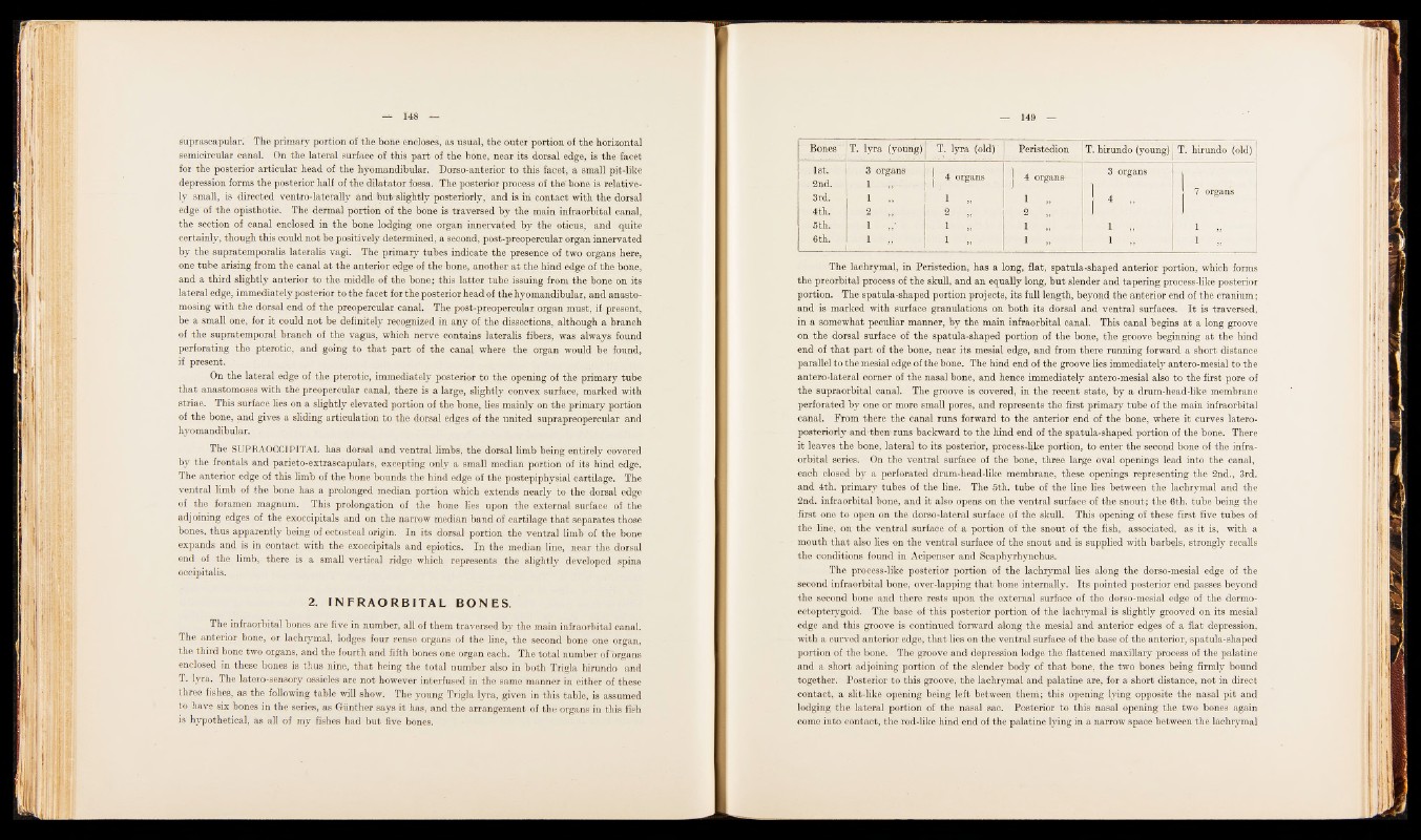
three fishes, as the following table will show. The young Trigla lyra, given in this table, is assumed
to have six bones in the series, as Giinther says it has, and the arrangement of the organs in this fish
is hypothetical, as all of my fishes had b u t five bones.
Bones ‘ T. lyra (young) T. lyra (old) Peristedion T. hirundo (young) T. hirundo (old)
1st. I . 3 organs
g§
1 4 organs
2nd. 1 ,,
3rd. 1
4th. 2
5th. 1
6th. 1
1
2 „
1
1
s|
4 organs-
1
2 „
1
1
3 organs
1
1 „
1
I 7 organs
1
1
The lachrymal, in Peristedion, has a long, flat, spatula-shaped anterior portion, which forms
th e preorbital process of th e skull, and an equally long, b u t slender and tapering process-like posterior
portion. The spatula-shaped portion projects, its full length, beyond the anterior end of the cranium;
and is marked with surface granulations on both its dorsal and ventral surfaces. I t is traversed,
in a somewhat peculiar manner, by the main infraorbital canal. This canal begins a t a long groove
on the dorsal surface of th e spatula-shaped portion of th e bone, the groove beginning a t th e hind
end of th a t p a rt of the bone, near its mesial edge, and from there running forward a short distance
parallel to the mesial edge of the bone. The hind end of the groove lies immediately antero-mesial to the
antero-lateral corner of the nasal bone, and hence immediately antero-mesial also to the first pore of
th e supraorbital canal. The groove is covered, in th e recent state, by a drum-head-like membrane
perforated by one or more small pores, and represents th e first primary tube of the main infraorbital
canal. From there th e canal runs forward to th e anterior end of th e bone, where it curves latero-
posteriorly - and th e n runs backward to the hind end of th e spatula-shaped portion of the bone. There
it leaves th e bone, lateral to its posterior, process4ike portion, to enter th e second bone of th e infraorbital
series. On the ventral surface of th e bone, three large oval openings lead into th e canal,
each closed by a perforated drum-head-like membrane, these openings representing the 2nd., 3rd.
and 4th. primary tubes of th e line. The 5th. tube of th e line lies between the lachrymal and the
2nd. infraorbital bone, and it also opens on th e ventral surface of the snout; the 6th. tube being the
first one to open on the dorso-lateral surface of th e skull. This opening of these first five tubes of
the line, on th e ventral surface of a portion of the snout of the fish, associated, as it is, with a
mouth th a t also lies on th e ventral surface of th e snout and is supplied with barbels, strongly recalls
th e conditions found in Acipenser and Scaphyrhynchus.
The process-like posterior portion of the lachrymal lies along the dorso-mesial edge of the
second infraorbital bone, over-lapping th a t bone internally. Its pointed posterior end passes beyond
the second bone and there rests upon the external surface of the dorso-mesial edge of the dermo-
ectopterygoid. The base of this posterior portion of the lachrymal is slightly grooved on its mesial
edge and this groove is continued forward along the mesial and anterior edges of a flat depression,
with a curved anterior edge, th a t lies on the ventral surface of the base of the anterior, spatula-shaped
portion of the bone. The groove and depression lodge the flattened maxillary process of the palatine
and a short adjoining portion of the slender body of th a t bone, the two bones being firmly bound
together. Posterior to this groove, the lachrymal and palatine are, for a short distance, not in direct
contact, a slit-like opening being left between them; this opening lying opposite the nasal pit and
lodging the lateral portion of the nasal sac. Posterior to this nasal opening the two bones again
come into contact, the rod-like hind end of the palatine lying in a narrow space between the lachrymal