the second, and differs from it in having only two apices to the middle ridge, and
only one internal cusp. The fourth molar is extremely minute, and presents only
two obscure cusps, one anterior and the other posterior.
The first incisors of the lower jaw project forwards and upwards, the terminal
fourth of their crowns being directed nearly upwards, and their tips are slightly
divergent, and their posterior surfaces more or less concave, but quite smooth.
The crown of the second incisor is the lowest of all the mandibular teeth, and is
elongately oblong when viewed externally, and is laterally compressed. The canine
is a little longer and more pyramidal, the crown being surmounted by a conical
point. The first molar has three external cusps, the first and last being nearly on the
same level, and the central being pyramidal and directed backwards. The latter
by its inner margin is connected by a short ridge to the anterior inner cusp, which
is immediately internal to it, rather sharply pointed, and nearly as high as itself.
The posterior internal cusp is opposite the last external cusp, but has on its outer
margin another short cusp between itself and that cusp, so that this tooth has six
cusps in all. The second molar is formed exactly like the first, but is much smaller.
The third molar is about one-third the size of the second, and the cingulum is well
marked along its outer side. I t developes two cusps, one small, anterior, and one
large, posterior and conical, and connected to the former by a short ridge more
internal than external to it.
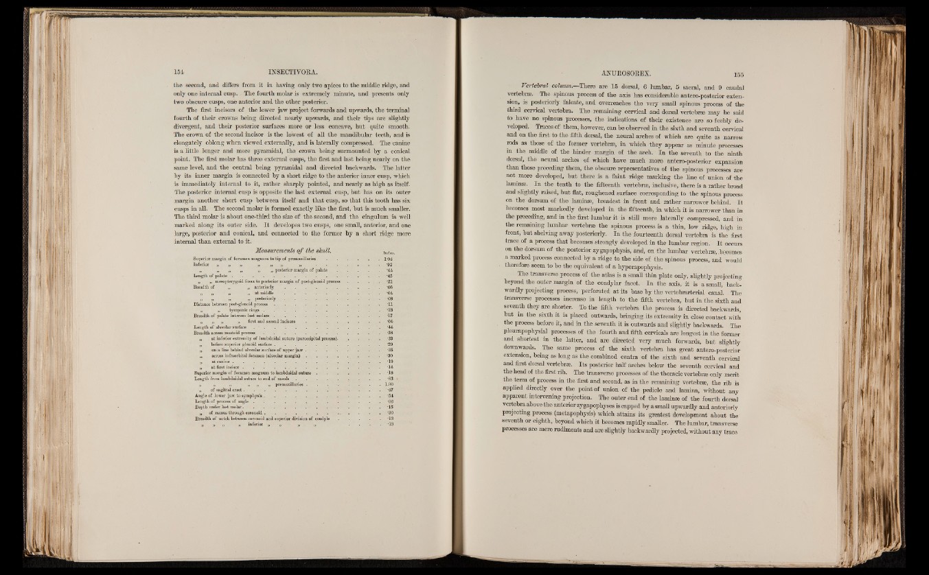
Measurements o f the skull. Inches.
Superior margin of foramen magnum to tip of premaxillaries . . . . . 1-04
Inferior „ „ ’ „ » „ „ „ . . . . .
„ „ „ „ „ „ posterior margin of palate
Length of palate ............................................................................... ..........
„ „ mesopterygoid fossa to posterior margin of post-glenoid process
Breadth of „ „ a n t e r i o r l y .....................................................................
„ „ „ „ at middle . . . . . . . .
Distance between post-glenoid process . . . . . . . . .
„ „ tympanic rings . . . . . . . . . .
Breadth o f palate between last molars . . . . . . . . .
„ „ „ „ first and second i n c i s o r s ..................................................
Length of alveolar surface ...................................................................................................
Breadth across mastoid process .........................................................................................
„ at inferior extremity of lambdoidal suture (paroccipital process).
„ before superior glenoid surface...............................................................................
„ on a line behind alveolar surface of upper jaw . . . . . .
„ across infraorbital foramen (alveolar m a r g in ) .................................................
„ at canine ............................................................................... ..........
„ at first incisor . . . . . . . . . . . . .
Superior margin of foramen magnum to lambdoidal s u t u r e ........................................
Length from lambdoidal suture to end o f n a s a l s ............................................................
„ „ | n n » premaxillaries..................................................
„ of sagittal crest. . . . . . . . . . . .
Angle of lower jaw to symphysis.........................................................................................
Length o f process of angle . . . . r . . . . .
Depth under last molar................................................. ...........................................................
„ of ramus through coronoid . . . . . . . . . .
Breadth of notch between coronoid and superior division of condyle
» » » » inferior „ „ „ 1 „ . -13
Vertebral column.—There are 15 dorsal, 6 lumbar, 5 sacral, and 9 caudal
vertebrae. The spinous process of the axis has considerable antero-posterior extension,
is posteriorly falcate, and overreaches the very small spinous process of the
third cervical vertebra. The remaining cervical and dorsal vertebrae may be said
to have no spinous processes, the indications of their existence are so feebly developed.
Traces of them, however, can be observed in the sixth and seventh cervical
and on the first to the fifth dorsal, the neural arches of which are quite as narrow
rods as those of the former vertebrae, in which they appear as minute processes
in the middle of the hinder margin of the arch. In the seventh to the ninth
dorsal, the neural arches of which have much more antero-posterior expansion
than those preceding them, the obscure representatives of the spinous processes are
not more developed, but there is a faint ridge marking the line of union of the
laminae. In the tenth to the fifteenth vertebrae, inclusive, there is a rather broad
and slightly raised, but flat, roughened surface corresponding to the spinous process
on the dorsum of the laminae, broadest in front and rather narrower behind. It
becomes most markedly developed in the fifteenth, in which it is narrower than in
the preceding, and in the first lumbar it is still more laterally compressed, and in
the remaining lumbar vertebrae the spinous process is a thin, low ridge, high in
front, but shelving away posteriorly. In the fourteenth dorsal vertebra is the first
trace of a process that becomes strongly developed in the lumbar region. I t occurs
on the dorsum of the posterior zygapophysis, and, on the lumbar vertebrae, becomes
a marked process connected by a ridge to the side of the spinous process, and would
therefore seem to be the equivalent of a hyperapophysis.
The transverse process of the atlas is a small thin plate only, slightly projecting
beyond the outer margin of the condylar facet. In the axis, it is a small, back-
wardly projecting process, perforated at its base by the vertebrarterial canal. . The
transverse processes increase in length to the fifth vertebra, but in the sixth and
seventh they are shorter. To the fifth vertebra the process is directed backwards,
but in the sixth it is placed outwards, bringing its extremity in close contact with
the process before it, and in the seventh it is outwards and slightly backwards. The
pleurapophysial processes of the fourth and fifth cervicals are longest in the former
and shortest in the latter, and are directed very much forwards, but slightly
downwards. The same process of the sixth vertebra has great antero-posterior
extension, being as long as the combined centra of. the sixth and seventh cervical
and first dorsal vertebrae. Its posterior half arches below the seventh cervical and
the head of the first rib. The transverse processes of the thoracic vertebrae only merit
the term of process in the first and second, as in the remaining vertebrae, the rib is
applied directly over the point of union of the pedicle and lamina, without any
apparent intervening projection. The outer end of the laminae of the fourth dorsal
vertebra above the anterior zygapophyses is capped by a small upwardly and anteriorly
projecting process (metapophysis) which attains its greatest development about the
seventh or eighth, beyond which it becomes rapidly smaller. The lumbar, transverse
processes are mere rudiments and are slightly backwardly projected, without any trace