nent canine is only appearing, the skull is considerably larger than that of the latter,
and also broader.
D. frenata, Gray, also differs from D. murina, M. and S., in its paler colour,
which is olive-brown, produced by the annulation of the hair. The under parts, also,
instead of being white as in the latter, are pale fawn. The black band from the snout
through the eye, behind which it expands backwards to the ear, where it ceases, is
less defined than in D. murina. There is a pale whitish, somewhat rufous streak or
band above this dark one, extending from the snout over the eye and ear, ceasing
behind the latter, but in D. murina the post-ocular portion expands into a large
white spot. There is also in both species a similar pale band below the black one,
but in neither is it expanded into a white spot, and it is more obscure in D. marma
than in D. frenata.
These two species stand as follows g n
D e n d r o g a l e m u r i n a , M. & S., Plate VII, figs. 1 8 and 1 9 , skull.
Eylogale murina, M. & S., Verhandl. (1889-44), i, p. 167, tab. 26, fig. 5 ; tab. 27, figs. 17 and 18.
Cladobates (Dendrogale, Gray) murinus, M. & S., Wagner, Schreber, Saugeth. (1855), vol. v, p. 528.
Habitat.—Borneo.
E e n d r o g a l e f r e n a t a , Gray, Plate VII, figs. 2 0 and 2 1 , skull.
Tupàia frenata, Gray, Ann. Mag. Nat. Hist., 1860, vol. vi, p. 217.
Habitat.—Camboja.
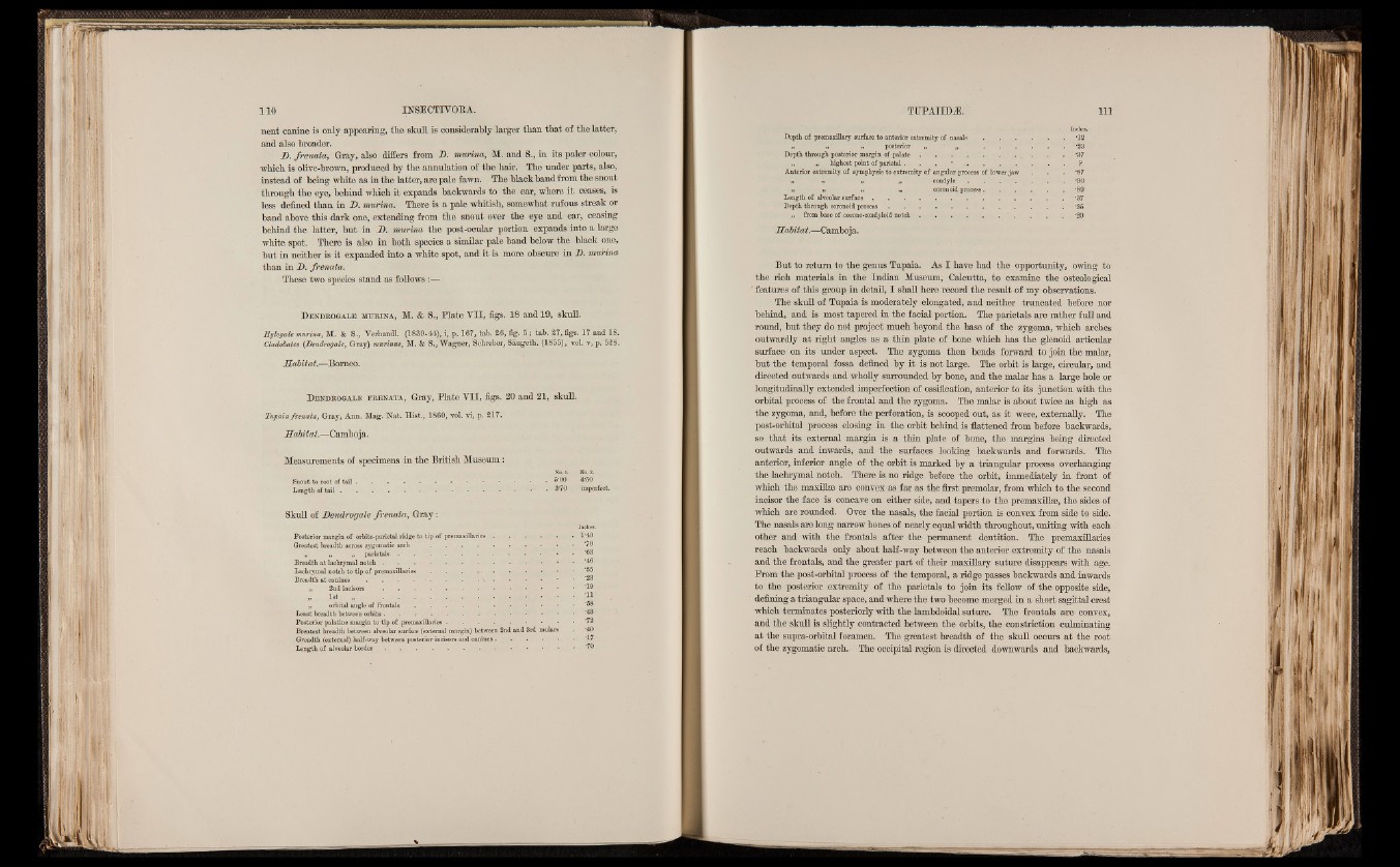
Measurements of specimens in the British Museum:
No. 1. No. 2.
Snout to root of t a i l .......................................................................................................................4'P®
Length o f t a i l • • . . • 3‘70 imperfect.
Skull of DencLrogale frenata, Gray:
Inches.
Posterior margin of orbito-parietal ridge to tip of premaxillaries . . . . . . 1’40
Greatest breadth across zygomatic a r c h ............................................................ >: ■ • ; •. <0
„ „ „ parietals . . . . . . • • ■ . . ’63
Breadth at lachrymal n o t c h ..................................................................... . • .- . ‘46
Lachrymal notch to tip o f premaxillaries . • . • ■ ■ • • - "5®
Breadth at canines . . . . • ■ • \ • • f . 2 3
i, 2nd incisors . . . . . . *1®
is t „ ;ii-
. , „ orbital angle o f f r o n t a l s ...................................................................................................
Least breadth between orbits . . . . • •
Posterior palatine margin to tip of premaxillaries...............................................................................
Breatest breadth between alveolar surface (external margin) between 2nd and 3rd molars . '40
Greadth (external) half-way between posterior incisors and canines....................................................... '17
Length o f alveolar border . . . . . .. • • • • "7P
Inches.
Depth of premaxillary surface to anterior extremity of n a s a l s ..........................................................'12
„ „ ,, posterior ■ „ ...................................................1 *23
Depth through posterior margin of p a l a t e .............................................................. . . . ‘37
„ „• . highest point o f parietal. . . .. P
Anterior extremity of symphysis to extremity of angular process of lower jaw . . '87
„ I. 1 • „ c o n d y l e .......................................................................... '90
i, » „ . „ coronoid process.................................................. ‘89
Length of alveolar surface , . . . . . . . . . . . *37
Depth through coronoid p r o c e s s ........................................................................................ . . *3S
„ from base o f .corono-condyloid n o t c h ........................................................... -20
Habitat.—Camboja.
But to return to the genus Tupaia. As I have had the opportunity, owing to
the rich materials in the Indian Museum, Calcutta, to examine the osteological
features of this group in detail, I shall here record the result of my observations.
The skull of Tupaia is moderately elongated, and neither truncated before nor
behind, and is most tapered in the facial portion. The parietals are rather full and
round, hut they do not project much beyond the base of the zygoma, which arches
outwardly at right angles as a thin plate of bone which has the glenoid articular
surface on its under aspect. The zygoma then bends forward to join the malar,
but the temporal fossa defined by it is not large. The orbit is large, circular, and
directed outwards and wholly surrounded by bone, and the malar has a large hole or
longitudinally extended imperfection of ossification, anterior to its junction with the
orbital process of the frontal and the zygoma. The malar is about twice as high as
the zygoma, and, before the perforation, is scooped out, as it were, externally. The
post-orbital process closing in the orbit behind is flattened from before backwards,
so that its external margin is a thin plate of bone, the margins being directed
outwards and inwards, and the surfaces lo o k in g backwards and forward?. The
anterior, inferior angle of the orbit is marked by a triangular process overhanging
the lachrymal notch. There is no ridge before the orbit, immediately in front of
which the maxillae are convex as far as the first premolar, from which to. the second
incisor the face is concave on either side, and tapers to the premaxillae, the sides of
which are rounded. Over the nasals, the facial portion is convex from side to side.
The nasals are long narrow bones of nearly equal width throughout, uniting with each
other and with the frontals after the permanent dentition. The premaxillaries
reach backwards only about half-way between the anterior extremity of the nasals
and the frontals, and the greater part of their maxillary suture disappears with age.
From the post-orbital process of the temporal, a ridge passes backwards and inwards
to the posterior extremity of the parietals to join its fellow of the opposite side,
defining a triangular space, and where the two become merged in a short sagittal crest
which terminates posteriorly with the lambdoidal suture. The frontals are convex,
and the skull is slightly contracted between the orbits, the constriction culminating
at the supra-orbitai foramen. The greatest breadth of the skull occurs at the root
of the zygomatic arch. The occipital region is directed downwards and backwards,