rounded process, concave from behind forwards in its hinder surface which forms
the anterior wall of the auditory meatus. In position it is considerably below the
upper border of the larger bulla, which arches inwardly over it and into the cavity
of which it projects, but leaving a fissure between its upper border and the arched
posterior margin of that bulla, the fissure being continuous with the cavities of the
larger bulla and tympanic. The cavity of the larger bulla is vertically oval and
looks inwards, the greater part of the hinder wall and the floor being formed by
the smaller bulla; this latter sends inwards forwards and upwards a curved ridge
from its base, a ledge-like floor to the cavity which is much antero-posteriorly
compressed and opening freely in the dried skull into the tympanic cavity. I t does
not occupy the whole of the bulla, for from the floor-ridge of the smaller bulla a
fine ridge is prolonged upwards, defining its inner limit, internal to which there
is a broad shallow groove running downwards behind its inner extremity where
the malleus is attached, viz., transverse to the longitudinal axis of the tympanic and
corresponding to the external bullate band. The inner surface of the cavity of the
bulla in the recent skull is external to the tympanic membrane. This membrane
is attached along the upper margin of the basal ridge (floor) of the smaller bulla,
and the fine ridge prolonged onwards from it along the inner margin of the cavity,
from the upper extremity of which it is reflected backwards to a ridge on the under
surface of the rough post-auditory process of the tympanic. Thence it continues
to the upper border of the tympanic, between that process and the smaller
bulla, and across the base of the latter internally, back to its basal ridge, but
above the opening into the cavity of this bulla, which latter is internal to
the tympanic, and a short, blind canal with a moderately wide orifice into the
tympanic cavity directed backwards. The inner surface of the cavity of the larger
bulla consists of a fine network of delicate ridges, the majority of which are
transverse, many of the intervening sulci being covered with very small pits.
A minute canal occurs in the floor of the cavity, and traversing the external
portion of the base of the smaller bulla, opens on the outside of the tympanic in
the lower end of the furrow, defining the posterior external border of the bullate
band.
I am indebted to Dr. Doran* for the following account of the ossicles of the ear of
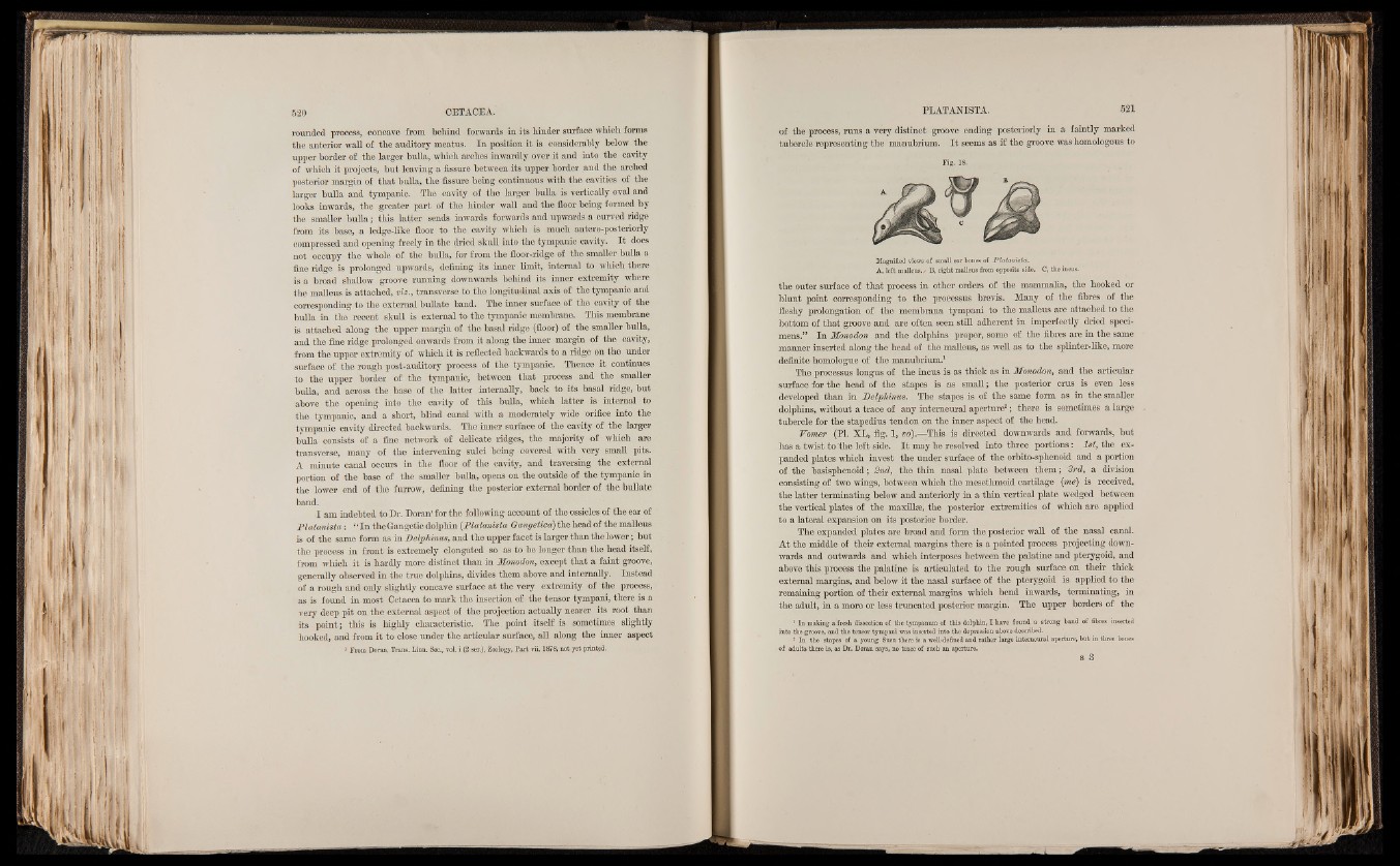
Flatmista: “ In the Gangetic dolphin (.Platanista Gcmgetica) the head of the malleus
is of the same form as in Delphmus, and the upper facet is larger than the lower; but
the process in front is extremely elongated so as to be longer than the head itself,
from which it is hardly more distinct than in Monodon, except that a faint groove,
generally observed in the true dolphins, divides them above and internally. Instead
of a rough and only slightly concave surface at the very extremity of the process,
as is found in most. Cetacea to mark the insertion of the tensor tympani, there is a
very deep pit on the external aspect of the projection actually nearer its root than
its point; this is highly characteristic. The point itself is sometimes slightly
hooked, and from it to close under the articular surface, all along the inner aspect
i From Doran, Trans. Linn. Soc., vol. i (2 ser.), Zoology, Part vii, 1878, not yet printgd.
of the process, runs a very distinct groove ending posteriorly in a faintly marked
tubercle representing the manubrium. I t seems as if the groove was homologous to
Fig. 18.
Magnified views of small ear bones of JPlatanista.
A, left malleus./ B, right malleus from opposite side. C, the incus.
the outer surface of that process in other orders of the mammalia, the hooked or
blunt point corresponding to the processus brevis. Many of the fibres of the
fleshy prolongation of the membrana tympani to the malleus are attached to the
bottom of that groove and are often seen still adherent in imperfectly dried specimens.”
In Monodon and the dolphins proper, some of the fibres are in the same
manner inserted along the head of the malleus, as well as to the splinter-like, more
definite homologue of the manubrium.1
The processus longus of the incus is as thick as in Monodon, and the articular
surface for the head of the stapes is as small; the posterior crus is even less
developed than in JDelphimts. The stapes is of the same form as in the smaller
dolphins, without a trace of any interneural aperture2; there is sometimes a large
tubercle for the stapedius tendon on the inner aspect of the head.
Vomer (PI. XL, fig. 1, vo).—This is directed downwards and forwards, but
has a twist to the left side. I t may be resolved into three portions: 1st, the expanded
plates which invest the under surface of the orbito-sphenoid and a portion
of the basisphenoid; 2nd, the thin nasal plate between them; 3rd, a division
consisting of two wings, between which the mesethmoid cartilage (me) is received,
the latter terminating below and anteriorly in a thin vertical plate wedged between
the vertical plates of the maxillae, the posterior extremities of which are applied
to a lateral expansion on its posterior border.
The expanded plates are broad and form the posterior wall of the nasal canal.
At the middle of their external margins there is a pointed process projecting downwards
and outwards and which interposes between the palatine and pterygoid, and
above this process the palatine is articulated to the rough surface on their thick
external margins, and below it the nasal surface of the pterygoid is applied to the
remaining portion of their external margins which bend inwards, terminating, in
the adult, in a more or less truncated posterior margin. The upper borders of the
1 In making a fresh dissection of the tympanum of this dolphin, I have found a strong band of fibres inserted
into the groove, and the tensor tympani was inserted into the depression above described.
2 In tbe stapes of a young Susu there is a well-defined and rather iarge interneural aperture, but in three bones
of adults there is, as Dr. Doran says, no trace of such an aperture.
s 3