compared with the shelving, almost concave frontal margin, to as far as the nipplelike
projections, and the fulness of the infero-frontal or orbito-fronta! parts are also
highly characteristic of the brain of Platanista. What is seen of the cerebellum in
side view, along with the several large nervous trunks, coincides in producing the
impression of height with vertical abruptness behind.
The occipital facies shows great steepness, or perpendicular shelving of the
parietal region, and equally oblique inwardly trending borders to the lips of the
longitudinal fissure, with a cutting away of the post-inner occipital lobes. The
cerebellum is less expansive than might be anticipated, though the nerve and
vascular channel impressions are apt to deceive in this respect.
Eye : its general appearance and structure.—(Pl. XXXVI, figs. 9 and 10.)|er;
Certainly one of the strongest peculiarities of the Gangetic dolphin (Platanista) is
the organ of vision. Frederick Cuvier remarks, “Les yeux sont fort petits, noirs, assez
profondément enchâssés dans leur orbite, et situés à environ deux pouces au-dessus
des coins de la bouche.” Eschricht states that the eyes are extraordinarily small in
diameter, only |L* line. I t may be called a blind Whale (according to him), for the
perforations for the optic nerve in the skull are only rudimentary. In describing
the young skull I shall afterwards refer to the optic foramen, (consult PI. XL,
fig. 1, op).
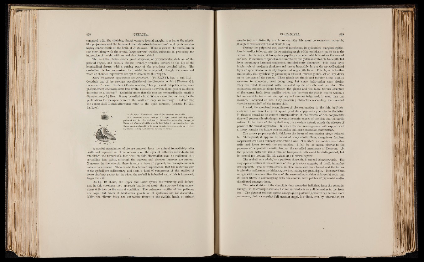
- F ig.-17.
A, the eye of P latan ista o f natural size, seen from the outside.
^ B, a horizontal section through the right eyeball including orbital
portion of skin, &c., of natural size ; f ., fatty cushion surrounding the eye ; fc ,
fibrous coat of same, with thin muscular layer below it ; m, muscular fibres ; fin,
fibrous membrane beneath ; s, the skin ; op, optic nerve ; c, eye-chamber ; i, iris ;
tel, internal eyelid ; el, el, external eyelids ; co, cornea.
A careful examination of the eye removed from the animal immediately after
death and repeated on three occasions on the eyes of different individuals, has
established the remarkable fact that, in this Mammalian eye, no rudiment of a
crystalline lens exists, although the aqueous and vitreous humours are present.
Moreover, in the choroid there is only a trace of pigment, and the optic nerve is
reduced to a thread. There is also this further imperfection, that the motor muscles
of the eyeball are rudimentary and form a kind of compressor of the cushion of
dense blubbery yellow fat, in which the .eyeball is imbedded and which is immensely
larger than it.
As fig. 10 shows, the upper and lower eyelids are relatively well defined,
and in this specimen they approach but do not meet; the aperture being narrow,
about O'20 inch in the natural condition. The cutaneous papillse of the palbebrse
are large; but traces of Meibomian glands or of eyelashes are not discernible.
Midst the fibrous fatty and connective tissues of the eyelids, bands of striated
muscles (m) are .distinctly visible so that the lids must be somewhat moveable,
though to what extent it is difficult to say.
Tracing the palpebral conjunctival membrane, its cylindrical marginal epithelium
is readily followed into the re-entering angle of the eyelid, as it passes on to the
cornea. In the angle,.it has quite a papillary character, which is lost on the corneal
surface. The comeal conjunctiva is nevertheless easily demonstrated, its free epithelial
layer assuming a flattened compressed stratified scaly character. This outer layer
is relatively of moderate thickness and passes insensibly into , a deeper well-defined
layer of spheroidal or vertically disposed oblong epithelium. This layer is further
and, notably distinguished by possessing a series of mucous glands which dip dowp
on to the face of the cornea. These glands are simple and tubular, a few slightly
racemose in character; most being long, but some intervening ones shorter.
They are filled throughout with nucleated epithelial cells and granules. The
submucous connective tissue between the glands and the more fibrous structure
of the cornea itself, form papillse which dip between the glands and in which, I
believe, could be traced minute capillary and nervous twigs, and, in more than one
instance, I observed an oval body possessing characters resembling the so-called
“ tactile corpuscles” of the human skin.
Indeed, the structural resemblances of the conjunctiva to the skin in Plata-
nista are close, save the great quantity of dark pigmentary matter in the latter.
I f . these observations be correct interpretations of the nature of, the conjunctiva,
they will go a considerable length towards the maintenance of the idea that the tactile
nature of . the front of the eyeball may, to a certain extent, supply the absence of
power in the visual apparatus. Whether further investigations will support such
a theory remains for future substantiation and more extensive examination.
The cornea proper equals in thickness the layers of conjunctiva above referred
to. Throughout, it appears to consist of wavy elastic fibres, elongate or fusiform
corpuscular cells, and ordinary connective tissue. The fibres are most dense internally
and looser towards the conjunctiva. I feel by no means clear as to the
presence of a posterior, elastic lamina, the so-called membrane of Demours. At
the junction. with the iris, a film of transparent cells could be distinguished, but
in none of my sections did this extend any distance beyond.
The eyeball, as a whole, has a pyriform shape, the blunt end being forwards. The
very open condition at the entrance of the optic nerve suggests, of itself, imperfect
development. The sclerotic coat is in close union with the choroid, and the former
is tolerably uniform in its thickness, nowhere having any great depth. Its outer fibres
mingle with the connective tissue of the surrounding cushion of large fat-cells, and
its inner fibres, in commingling with the choroid, have patches of pigmental matter
distributed amongst them.
The outer division of the choroid is thus somewhat indistinct from the sclerotic,
though, in, microscopic sections, the retinal border is as well defined as in the fresh
, eye. The pigment cells are sparse, except quite posteriorly, where they become more
numerous; but a somewhat full vascular supply is evident, even by observation on