mucous membrane is greyish, and the only traces of rugae that can be detected are a
few isolated patches of lobular processes, of the membrane, which is generally smooth,
but shows the persistent folds. In the last condition, the glandular surfaces are
most easily detected. I t is sparsely covered with a number of reddish spots of varied
size and form, in which the mucous membrane is coarser than it is generally, but no
special gland openings can be detected in these localities. A remarkable glandular
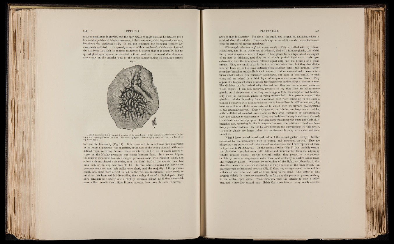
area occurs on the anterior wall of the cavity almost facing the opening common
Fig. 14.
M
(W7®l
B
A.
A sketch (natural size) of the surface o f a portion of the second cavity of the stomach of Platanista at the part
where the “ cup-shaped bodies” are found. The subsidiary figure B shows a highly magnified view of a few of the
cup-shaped bodies.
to it and the first cavity (Eig. 14). I t is irregular in form and is*at once discernible
by its rough appearance; the rugosities, in the case of the young stomach with well-
defined rugae, occurring between these structures; and in the stomachs devoid of
rugae, on the lobular processes, but chiefly between them. In a young dolphin
the mucous membrane has raised rugged processes, some with rounded heads, and
others with cup-shaped extremities, as if the distal half of the rounded head had
been lost, or the cup had lost its lid. In two adults nothing but cup-shaped
processes remained, and their stalks were short, and the majority of the processes
small, and some were almost buried in the mucous membrane. They recall to
mind, in their form and definite outline, the sucking discs of a Cephalopod. They
have considerable tenacity and a slightly brownish colour, as if they were chiti-
nous in their constitution. Such little cups,—and there must be some hundreds,—
are 0'05 inch in diameter. The rim of the cup is not its greatest, diameter, which is
attained about the middle. These single cups in the adult are also connected to each
other by strands of mucous membrane.
Microscopic characters o f the second cavity.—This is clothed with cylindrical
epithelium, and in its whole extent is densely clad with tubular glands, into which
the cylindrical epithelium is prolonged. These glands form a layer about one-eighth
of an inch in thickness, and they are so closely packed together at their open
extremities that the interspaces between equal only half the breadth of a gland
tubule. They are simple tubes in the first half of their extent, but they then divide
into two branches, and in some instances bend suddenly before the division. These
secondary branches rapidly diminish in capacity, and are soon reduced to narrow tortuous
tubules which run vertically downwards, but more or less parallel to each
other, and are lodged in a thick layer of corpusculated connective tissue. They
appear also to give off other branches like themselves maintaining a similar course.
The divisions can be undoubtedly observed, but they are not so numerous as one
would expect. I am not, however, prepared to say that they are all racemose
glands, but if simple ones occur, they would appear to be the exception and to differ
only from the compound glands in being unbranched. I t appears to me as if the
glandular tubules, depending from a common duct were bound up in one sheath,
because I observed even as many as from two to four orifices, in oblique section, lying
together as if in a cellular mass, external to which were the upward prolongations
of the muscular mucosa. These cells around the tubules are large ovoid vesicles,
with well-defined rounded nuclei, and, as they were unstained by hsematoxylon,
they are difficult to demonstrate. They are doubtless the peptic cells seen through
the delicate membrana propria. The cylindrical cells lining the ducts and their chief
branches, and occurring in the interspaces between the orifices of the ducts, have
finely granular contents. In the hollows between the convolutions of this cavity,
the peptic glands are larger tubes than on the convolutions* but shorter and more
branched.
What I have termed cup-shaped bodies of this second gastric cavity I further
examined by the microscope, both in vertical and horizontal section. They are
altogether very peculiar and quite anomalous structures, and I have represented them
in figs. 1 and 2, PI. XXXVII. In the vertical section (Eig. 1) they partially occupy
the glandular layer, but seem quite distinct and circumscribed from the adjoining
tubular mucous glands. In the vertical section, they present a homogeneous
or faintly granular egg-shaped outer area, and centrally a darker ovoid mass,
also vertically- placed. Whether by refraction of the light, or otherwise, in this
view there seems to be a central band in the long direction of the inner object. In
the transverse or horizontal sections (Eig. 2) these cup or egg-shaped bodies exhibit
a thick circular outer wall, with an inner lining to the same. This latter is bent
inwards chiefly in three, or occasionally in four, angular pieces projecting midway
to the central open space. They, therefore, cause the interior to have a trifoil
area, and where they almost meet divide the space into as many nearly circular