These recall to mind the moniliform vessel of Orca and the sac-like dilatations
of the arteries on the stomach of Orcella. In other instances the venous channels
send off a diverticulum or side chamber (fig. 7), which occasionally is connected by
a narrowed orifice with a subsidiary sac.
In a partially transparent transverse section of the umbilical cord (PI. XXXVI,
fig. 11), the arrangement of the two main arteries and two veins is shown, and
besides, marginally and between the latter, a large vacuity, a probable remnant of
the urachus. Both arteries and veins have relatively thick and dense walls. The
urachus cavity is much thinner-walled, and interiorly I made out the remains
of minute and short papillae with an epithelial covering. Towards the peripheral
margin of the cord the gelatine of Wharton is loose-meshed, and traversed by narrow
ellipsoidal channels. Although these approach the superficial glandular bodies, they
nevertheless do not seem to have any connection with them.
Foetus (PI. XXXI, fig. 1, and PI. XXXII, fig. 4).—The mouth of the fcetus
was only partially closed, hut its openness was probably a post-mortem result,
as the palatal surface had a longitudinal mucous ridge corresponding to a deep
groove in the middle of the flat prelingual surface. The outer aspect of both
jaws was raised into well-marked vertical ridges, and increasing in size from
behind forwards and terminating in marginal papillary eminences, through which
the sharp teeth could he.felt; and on the right side there were eighteen such
columns more or less visible on both jaws, and sixteen ,on the upper and
eighteen on the lower on the left side. The angles of the mouth and the
blow-hole were filled with a consistent grumous substance which appeared to
be cast and disintegrated epithelium, as a few broken nucleated epithelial cells
could be detected in it under the microscope. The pectoral flippers were laid
backwards against the sides of the body, and the free margin of the right was
slightly folded on itself. The dorsal fin was bent to the left side. The left caudal was
folded inwards against the under surface of the right flipper, while its tip was bent
over its own upper surface; the tip only of the right caudal flipper being folded in
a manner similar to its fellow. In the two fcetuses which have come under my
observation the bodies have been marked by corresponding folds. Among the most
prominent of these is a deep fold which marks the position of the neck, occurring
half-way between the angle of the mouth and the pectoral flipper. I t is deepest on
the ventral surface and extends up the side to nearly on a level with the eye. Two
other somewhat similar folds occur, one before and the other behind the anus, but
as they are due to the downward and forward bending of the caudal portion of the
animal they disappear shortly after birth. On the dorsal surface there are four large
folds which have been alike in both fcetuses: a shallow transverse fold shortly
behind the blow-hole and corresponding in position to the gular fold; a second
somewhat double fold stretching between the pectoral flippers; and another occurring
at a third of the distance between the latter structures and the tip of the dorsal
flipper; and a fourth half-way between the third fold and the dorsal fin. Behind
the dorsal fin there are seven to eight short transverse folds across the rather sharp
ridge of the back, and these remain a long time persistent. Besides these folds
the skin generally is covered with wavy, transverse wrinkles, which however
disappear in adult age in a healthy dolphin. The upper surface of the snout
(PI. XXXI, fig. 1, p.) is covered by scattered, rather strong, palish yellow bristles,
and the under surface of the lower jaw with twenty-six similar hairs. I have not
been able to detect hairs in any other part of the body and there is no whisker or
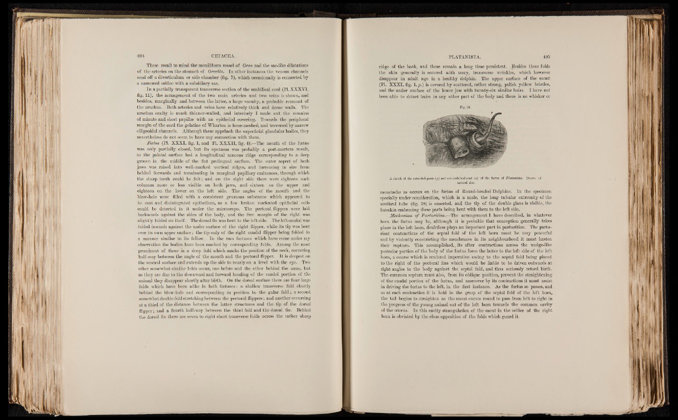
Fig. 18.
A sketch of the extended penis (p) and cut umbilical cord («) of the fcetus of Platanista. Drawn of
natural size.
moustache as occurs on the foetus of Bound-headed Dolphins. In the specimen
specially under consideration, which is a male, the long tubular extremity of the
urethral tube (fig. 18) is exserted, and the tip of the double glans is visible, the
foreskin embracing these parts being bent with them to the left side.
Mechanism of Parturition.—The arrangement I have described, in whatever
horn the foetus may be, although it is probable that conception generally takes
place in the left horn, doubtless plays an important part in parturition. The parturient
contractions of the septal fold of the left horn must be very powerful
and by violently constricting the membranes in its neighbourhood it must hasten
their rupture. This accomplished,, its after contractions across the wedge-like
posterior portion of the body of the foetus force the latter to the left side of the left
horn, a course which is rendered imperative owing to the septal fold being placed
to the right of the pectoral fins which would be liable to be driven outwards at
right angles to the body against the septal fold, and thus seriously retard birth.
The common septum must also, from its oblique position, prevent the straightening
of the caudal portion of the fcetus, and moreover by its contractions it must assist
in driving the fcetus to the left, in the first instance. As the foetus so passes, and
as at each contraction it is held in the grasp of the septal fold of the left horn,
the tail begins to straighten as the snout curves round to pass from left to right in
the progress of the young animal out of the left horn towards the common cavity
of the uterus. In this cavity strangulation of the snout in the orifice of the right
horn is obviated by the close apposition of the folds which guard it.