o N E N
TABLE PAGXV.
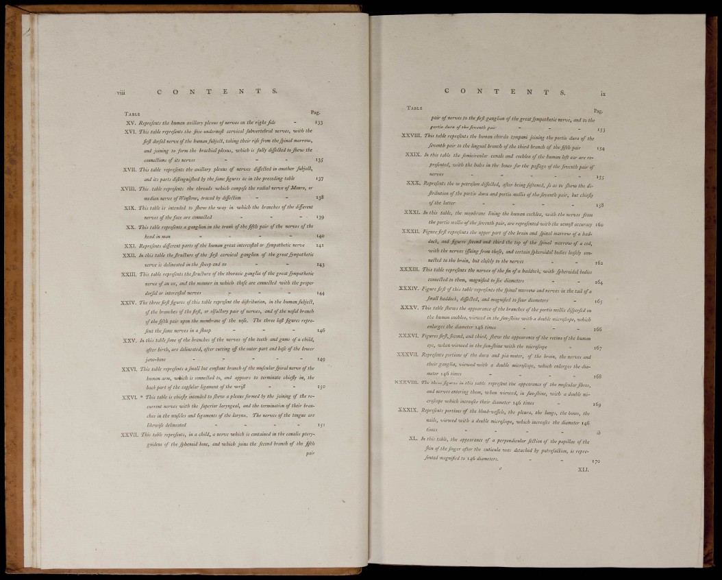
Reprejents the human axillary plexus of nemes on the right ftde - 133
XVI. This table repreßnts the five undennoß cervical fubvertebral nerves, -with the
firfl dorfal nerve of the human fubjeä, taking their rife from thefpinal marro-w,
and Joining to form the brachial plexus, ivhich is fully differed to Jhevj the .
connexions of its nerves - - - 13^
XVII. This table reprefents the axillary plexus of nerves diffeEied in another fubjeiî,
and its parts dißinguißed by the fame figures as in the preceding table 137
XVIII, This table reprefents the threads -which compofe the radial nerve of Monro, or
median nerve of Winßow, traced by dißBion - - 138
XIX. This table is intended to ß^evj the -way in ivhich the branches of the different
nerves of the face are conneiîed - - - 139
XX. This table reprefents a ganglion in the trunk of the fifih pair of the nerves of the
head in man - - - - 140
XXI. Reprefents different parts of the human great intercoßal or fympathetic nerve 141
XXII. In this table the ßruäure of the firß cervical ganglion of the great fympathetic
nerve is delineated in the ßeep and OX - - 143
XXIII. This table reprefents theßrußure of the thoracic ganglia of the great fympathetic
nerve of an ox, and the manner in which thefe are conneiled -with the proper
dorfal or int-ercoßal nerves r ~ " ^44
XXIV. The three firß figures of this table reprefent the difiribution, in the human fuhjeEl,
of the branches of the firß, or olfaäory pair of nerves, and of the nafal branch
of the fifth pair upon the membrane of the nofe. The three laß figures reprefent
the fame nerves in a ßeep - - - 146
XXV. In this table fome of the branches of the nerves of the teeth and gums of a child,
after birth, are delineated, after cutting off the outer part and bafc of the hiver
jaiv'bone - - _ - _ 14g
XXVI. This table reprefents a fmall but confiant branch of the mufcular fpiral nerve of the
human arm, ivkich is conneäed to, and appears to terminate chiefiy in, the
back part of the capfular ligament of the ivrifi - - 150
XXVI. * This table is chiefiy intended to fiieiu a plexus formed by the Joining of the recurrent
nerves -with the fuperior laryngeal, and the termination of their branches
in the mufcles and ligaments of the larynx. The nerves of the tongue are
likeivife delineated - - - - 15'
XXVII. This table reprefents, in a child, a nerve ivhich is contained in the canalis pterygoideus
of the fphenoid bone, and ivhich joins the fécond branch of the fifth
pair
O N
TABLE
Pag.
« of the great fympathetic nerve, and to the
X X V I I I . ;
X X X . .
X X X I .
X X X U I .
pair of nerves,to t
portio dura of the feventh pair - - _
. This table reprefents the human chorda tympani Joining the portio dura of the
feventh pair to the lingual branch of the third branch of the fifth pair 154
.. In this table the femicircular canals and cochlea of the human left ear are reprefented,
vjith the holes in the bones for the paffage of the feventh pair of
nerves
. Reprefents the os petrofum differed, afier being foftened, fo as to Pew the difiribution
of the portio dura and portio mollis of the feventh pair, but chiefiy
of the latter - _ _ _
. In this table, the membrane lining the human cochlea, with the nerves from
the portio mollis of the feventh pair, are reprefented with the utmofi accuracy 160
. Figure firfi reprefents the npper part of the brain and fpinal marrow of a haddock,
and figures fécond and third the top of the fpinal
marríTM of a cod^
•wlli the ncraei ¡JJUingfrom thefe, and certain fphenidal todies loofely con-
1te¿ied to the brain, but chfely to the nervei - _ jgj
. This table represents the nerms tf the fin of a haddock, -with ffheroidal bodies
conneEied to them, magnified to Jtx diameters - _ ¡g^
XXXIV. Figure firjl of this table reprefents the fpinal nrarrmt and nerves in the tail of a
fmall haddock, dijeñed, and magnified to four diameters - j
XXXV. This table fhews the appearance of the branches of the portio mollis dfperfed on
the human cochlea, „ienned in the fiin-fhine -with a doable micro/cope, -which
enlarges the diameter 146 times _ - - 166
XXXVI. Figures firfl, feeond, and third, fjie-ui the appearance of the retina of the human
eye, -when -viewed in thcfuu-fhine -with the microfeope - 167
XXXVII. Reprefents portions of the dura and pia mater, of the brain, the •nerves and
their ganglia, -viewed -uiith a double microfcope, ivhich enlarges the diameter
I times ~ _ - 168
XXXVIIL. ne three figures in this table reprefent the appearance of the mufcular fibres,
and nerves entering them, -when vietved, in fun-fhiue, -with a double microfcope
-which iiicreafei their diameter 146 times -
orefent, portions of the blood-veffels, the pleura, the lungs, the bones, the
nails,times -viewed -wi-t h a double m_i crofcope, -which increafes the diameter 14S ,, lb
L. lu this table, the appearance of a perpendicular feñion of the papillae of the
Jkln of the finger after the eutieula ivas detached by putrefañion, is reprefented
magnified to il^i diameters. - .
XXXIX. Repn,
B