^ l l l
1
I X B . xxix
•si 1 - »
E X P L A N A T I O N OF T H E TABLES. 155
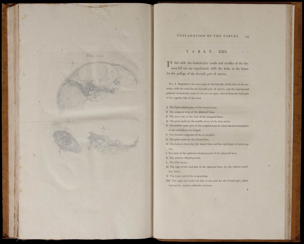
T A B L E XXIX.
J N this table the femiclrcular canals and cochlea of the human
left ear are reprefented, with the holes in the bones
for the paiTage of the feventh pair of nerves.
FIG. I. Reprefents the inner part of the left fide of the bafe of the cranium,
with the canal for the feventh pair of nerves, and the fuperior and
poilerior fcmicircular canals of the ear cut open, viewed from the back part
of the oppofite fide of the head.
A The fupra orbital plate of the frontal bone.
B The temporal wing of the fphenoid bone.
C The inner fide of the fcale of the temporal bone.
D The print made by the middle artery of the dura mater.
E The hollow under part of the occipital bone in which the left hemifphere
of the cerebellum was lodged.
F The foramen magnum of the os occipitis.
G The print made by the lateral finus.
H The hole at which the left lateral finus and the eighth pair of nerves go
out.
I The back of the pofterior chnoid procefs of the fphenoid bone.
K The anterior clinoid procefs.
L The fella turcica.
M The edge of the oval hole of the fphenoid bone for the inferior maxillary
nerve.
N The inner end of the os petrofum.
0 0 The upper and under cut fides of the canal for the feventh pair, called,
improperly, meatus auditorius internus.
P