E X P L A N A T I O N O F T H E T A B L E S .
T A B L E III.
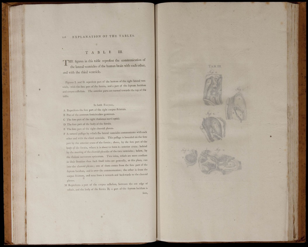
" "T^HE figures in tliis table reprefent the communication of
the lateral ventricles of the human brain with each otlier,
and with the third ventricle.
Figures I. and II. reprefent part of the bottom of the right lateral ventvicle°
with the fove part of the fornix, and a part of the feptum lucidum
and corpus callofum. Tlic anterior parts are turned towards the top of the
table.
In both FIGURES,
A Repvefents tlie fore part of the right corpus ftriatum.
B Part of the centrum femicircukre geminum.
C The fore part of the right thalamus nervi optici.
D The fore part of the body of the fornix.
E The fore part of the right choroid plexus.
F A natural palTage by which the lateral ventricles communicate with each
other and with the third ventricle. This paffagc is bounded on the fore
part by the anterior crura of the fornix; above, by the fore part of the
body of the fornix, where it is about to form its anterior crura; behind
by the meeting of the choroid plexufes of the two ventricles; below, by
the thalami nervorum opticorum. Two veins, which are more conftant
in their f.tuation than fuch fmall veins are generally, at this place, run
into the choroid plexus ; one of them comes from the fore part of the
feptum lucidum, and is over the communication; the other is from the
corpus fbriatum, and runs from it inwards and backwards to the choroid
plexus. =
M Reprefents a part of the corpus callofum, between the cut edge of
which, and the body of the fornix D, a part of the feptum lucidum is
feen.
H 111
' / O
J C t - i M
r ' - X i '
• f J #