Such a high ratio is again characteristic of this body plan.
The limbs were fin-shaped, with zeugopodials retaining a
shaft but not as elongated as in the basal plan. The carpals
are rectangular or squarish, being tightly packed, but are
distinct from the metacarpals and phalanges that retain
shafts.
45 (±5) presacral vertebrae. The trunk vertebral centra are
discoidal, with height/length ratios of about 2.5 near the
pelvic girdle. There is some increase of centra height posterior
to the caudal peak (i.e., tailbend), although not as
extensively as in the mixosaurian plan. The metacarpals
and phalanges lack shafts, and the carpals are closely
packed.
Parvipelvian plan
(Fig. 68F-G)
The parvipelvian plan is known from numerous complete
skeletons, and is the best known of the four body plans.
Most characteristically, the pelvic girdle is remarkably reduced
anteroposteriorly. The caudal peak has been modified
to form a tailbend, supporting a lunate caudal fin. A
dorsal fin is present at least in some forms. There are about
Californosaurus
(Fig. 68E)
This genus probably represents a body plan that is basal to
the parvipelvian plan. The presacral count is about 45 and
there appears to be a tailbend, but the pelvic girdle is still
moderately large.
Review of Body Part Evolution
Skull
(Fig. 69)
The cranial bones in ichthyopterygians overlap one another
extensively. This sometimes leads to misinterpretation
of sutural lines, because apparent sutures move when the
surface bone breaks off, exposing the underlying element.
Also, variation in bone growth among and within individuals
could move the suture extensively for similar reasons.
In addition, certain elements, such as the jugal, may be
partially disarticulated, and this could lead to misinterpretation
of suture lines. Therefore, attention should be paid
to preservational factors as well as individual and ontogenetic
variation when interpreting ichthyopterygian skulls,
and whether or not one bone misses contact with another
by a small amount should not be taken seriously in phylogenetic
discussions.
The skulls of all ichthyosaurs share a single basic plan.
The snout is narrow and elongated, with the external nares
located proximally rather than at its rostral tip. The orbit is
large, with a narrow, rod-like jugal forming its ventral
margin. The scleral ring is also large (but not always preserved).
There is a pair of supratemporal fenestrae, but no
definitive infratemporal fenestrae. A pineal foramen is always
present. Ichthyopterygian cranial structure diversified
within this basic framework.
The pineal foramen is enclosed between the anterior
processes of the parietals in basal forms, at least in dorsal
view. It is possible that the frontals made a contribution to
the foramen on the ventral aspect of the skull roof. The
anterior parietal processes became reduced in the stem
plan to form a parietal fork (Motani, 1999C), and thus the
pineal foramen was located on the suture of the parietal
and frontal, being enclosed within the fork. The regression
of the parietal along the medial line further proceeded in
the Parvipelvia, where the pineal foramen is sometimes
entirely enclosed between the frontals.
The parietal also went through a remarkable modification
posteriorly. In basal forms and Cymbospondylus, the
posterolateral (supratemporal) process of the parietal was
long and narrow, and the parietal ridge, a structure unique
to derived ichthyosaurs, was absent. This latter structure
appeared in Mixosaurus and persisted in parvipelvians,
although its presence has yet to be confirmed for shasta-
saurines. The supratemporal process was shortened to
various degrees in these forms.
Another important feature of the skull roof is the anterior
terrace of the supratemporal fenestra, which is the
depressed area of the skull roof along the anterior margin
of the supratemporal fenestra. The terrace appears as if it
were sculpted on the skull roof, with its anterior limit
clearly marked by a parabolic line. The presence of this
structure is plesiomorphic for ichthyopterygians. Sutural
lines are difficult to follow on the terrace, but it appears
that three bones, namely the parietal, frontal, and postfrontal,
were involved in the formation of this structure in basal
ichthyosaurs (Motani et al., 1998; Motani, 2000A). The
terrace became smaller in the stem ichthyosaurs, as the
frontal became eliminated from the area. Most parvipelvians
lost this structure, but at least one specimen of Lepto-
nectes (ROM 30127) and another of an undescribed new
species from the Lower Lias (BMNH R3000) retained it.
The terrace is clearly defined only on the postfrontal in
these specimens, but it should also be considered that the
postfrontal is remarkably enlarged in parvipelvians compared
to the more basal forms. A reverse trend is seen in
mixosaurians: the terrace was remarkably expanded in
these forms, almost reaching the external naris anteriorly,
and this large terrace is one of the synapomorphies for the
Mixosauria (Motani, 1999B, 1999C). The functional significance
of these enlarged terraces is unclear. One possibility
is that they accommodated salt glands in the expanded
anterior region; the area corresponds to the position of salt
glands in extant marine iguanas (Dunson, 1976).
The basic configuration of the cheek region remained
unchanged, although there were variations in the relative
extent of areas occupied by the bones involved (viz., the
supratemporal, squamosal, quadratojugal, jugal, postorbital,
and postfrontal). These six elements were almost
always present, except for the squamosal that has been
reported to be absent in Ichthyosaurus (McGowah 1973B)
and Platypterygius (Romer, 1968) (see discussion above).
The contact between the jugal and quadratojugal was absent
in basal ichthyosaurs, but present in derived forms.
Plesiomorphically, the postfrontal had a posterior process
that overlapped the postorbital, but the process became
reduced and eventually disappeared in derived forms.
The external nares were located close to the sagittal
plane in basal ichthyopterygians, with a narrow bar formed
by the nasal and premaxilla separating the paired openings.
They faced dorsolaterally in these forms, suggesting
that the animals may have been able to breathe while
largely submerged in the water. In the stem, mixosaurian,
and parvipelvian skulls, the nasal in the narial area is much
thickened and elevated, displacing the nares toward both
sides of the skull. These laterally facing nares would have
required the animals to emerge a large part of their heads
to break the water surface to breathe.
Dentition
The tooth crowns are mostly conical, but there are variations
and exceptions. Rounded crowns are known in the
posterior part of the jaw in Chaohusaurus, Grippia, Mixosaurus,
and Tholodus, but this feature is probably not synapo-
morphic among the four. The posterior teeth of Grippia and
Chaohusaurus are wide labiolingually (Motani, 1997B), as
in the conical posterior teeth of Utatsusaurus (Motani,
1996), whereas those of the other two genera are long
mesiodistally (i.e., labiolingually compressed) (Merriam,
1910; Peyer, 1939). Durophagy has been proposed for the
four genera, but there is no direct evidence of such feeding
habits (e.g., stomach contents). The jaws of Grippia, Chaohusaurus,
and M. cornalianus are possibly too slender for
durophagy, and their small teeth may have been used for
grasping prey, rather than crushing it (Motani, 1997A,
1997B). The teeth of M.fraasi and M. nordenskioeldii as well
as Tholodus clearly show adaptation for shearing, so they
may have been truly durophagous. The rounded tooth
crowns are associated with the occurrence of multiple
maxillary tooth rows, except in M. cornalianus.
The possession of unusually small teeth is characteristic
for some basal ichthyosaurs (e.g., Motani, 1996,1997B).
The maximum dimension of the crown is smaller than five
percent of the skull width in these teeth, a condition otherwise
unknown in marine reptiles (Massare, 1987). It is
possible that basal ichthyosaurs had a different diet from
all other known marine reptiles.
Another remarkable departure of the crown shape
from the typical conical design is labiolingual flattening,
resulting in the presence of two cutting edges, mesially
and distally. This type of tooth crown is only known in
large ichthyosaurs. The most remarkable teeth are those of
Himalayasaurus, which do not appear very ichthyosaurian
because of the extreme flattening, as well as some swelling,
of their tooth crowns (Motani et al., 1999). Temnodontosau-
rus is also known to have somewhat flattened crowns,
although these coexist with the typical conical type (McGowan,
1974A, 1979B).
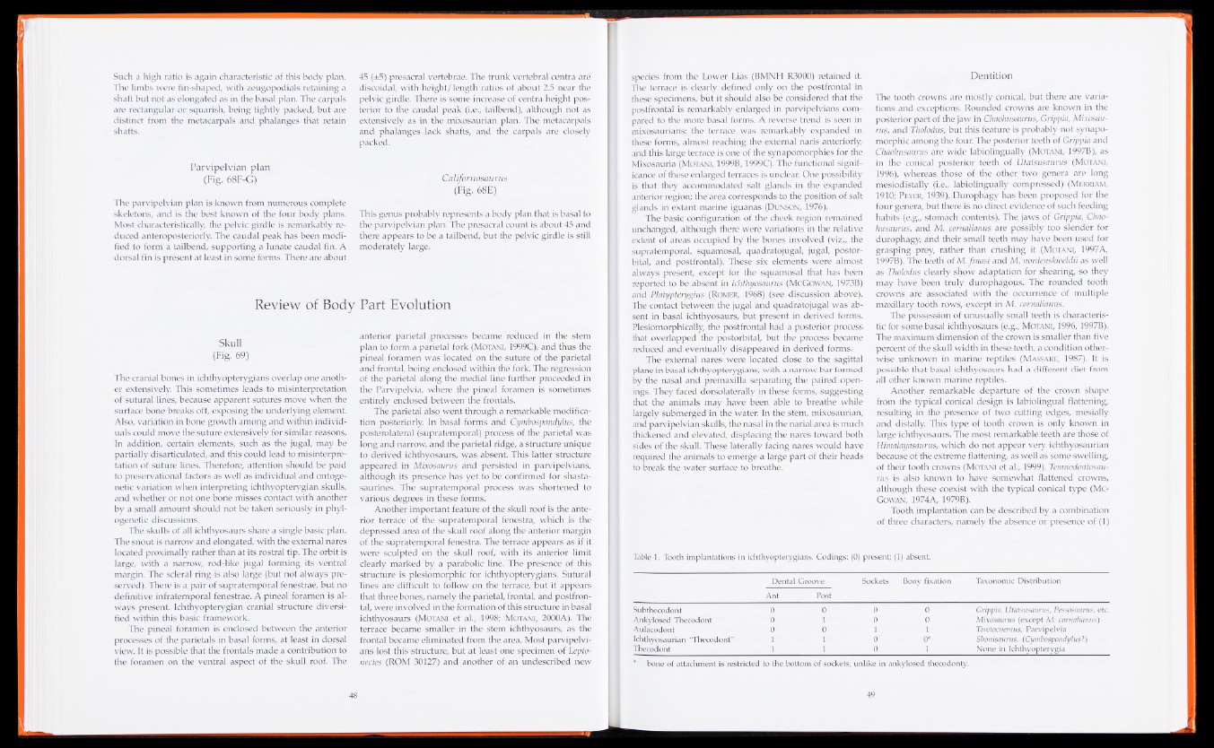
Tooth implantation can be described by a combination
of three characters, namely the absence or presence of (1)
Table 1. Tooth implantations in ichthyopterygians. Codings: (0) present; (1) absent.
Dental Groove Sockets Bony fixation Taxonomic Distribution
Ant Post
Subthecodont 0 0 0 0 Grippia, Utatsusaurus, Pessosaurus, etc.
Ankylosed Thecodont 0 1 0 0 Mixosaurus (except M. cornalianus)
Aulacodont 0 0 1 1 Toretocnemus, Parvipelvia
Ichthyosaurian “Thecodont” 1 1 0 0* Shonisaurus, (Cymbospondylus?)
Thecodont 1 1 0 1 None in Ichthyopterygia
* bone of attachment is restricted to the bottom of sockets, unlike in ankylosed thecodonty.