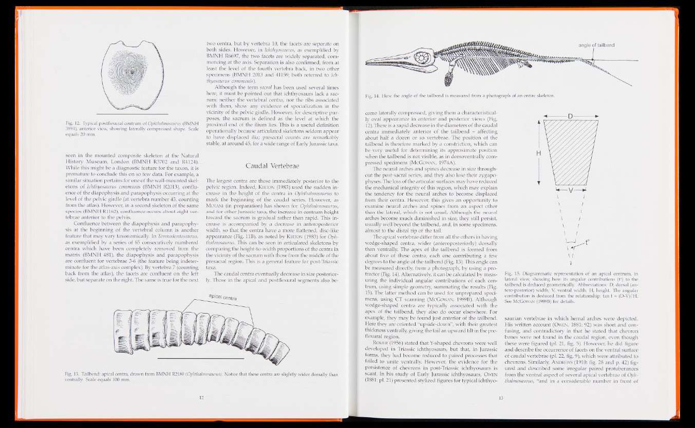
Fig. 12. Typical postflexural centrum of Ophthalmosaurus (BMNH
3894), anterior view, showing laterally compressed shape. Scale
equals 20 mm.
seen in the mounted composite skeleton at the Natural
History Museum, London (BMNH R3702 and R4124).
While this might be a diagnostic feature for the taxon, it is
premature to conclude this on so few data. For example, a
similar situation pertains for one of the wall-mounted skeletons
of Ichthyosaurus communis (BMNH R2013), confluence
of the diapophysis and parapophysis occurring at the
level of the pelvic girdle (at vertebra number 43, counting
from the atlas). However, in a second skeleton of the same
species (BMNH R1162), confluence occurs about eight vertebrae
anterior to the pelvis.
Confluence between the diapophysis and parapophysis
at the beginning of the vertebral column is another
feature that may vary taxonomically. In Temnodontosaurus,
as exemplified by a series of 65 consecutively numbered
centra which have been completely removed from the
matrix (BMNH 481), the diapophysis and parapophysis
are confluent for vertebrae 3-6 (the feature being indeterminate
for the atlas-axis complex). By vertebra 7 (counting
back from the atlas), the facets are confluent on the left
side, but separate on the right. The same is true for the next
two centra, but by vertebra 10, the facets are separate on
both sides. However, in Ichthyosaurus, as exemplified by
BMNH R6697, the two facets are widely separated, commencing
at the axis. Separation is also confirmed, from at
least the level of the fourth vertebra back, in two other
specimens (BMNH 2013 and 41159; both referred to Ichthyosaurus
communis).
Although the term sacral has been used several times
here, it must be pointed out that ichthyosaurs lack a sacrum:
neither the vertebral centra, nor the ribs associated
with them, show any evidence of specialization in the
vicinity of the pelvic girdle. However, for descriptive purposes,
the sacrum is defined as the level at which the
proximal end of the ilium lies. This is a useful definition
operationally because articulated skeletons seldom appear
to have displaced ilia; presacral counts are remarkably
stable, at around 45, for a wide range of Early Jurassic taxa.
Caudal Vertebrae
The largest centra are those immediately posterior to the
pelvic region. Indeed, Kirton (1983) used the sudden increase
in the height of the centra in Ophthalmosaurus to
mark the beginning of the caudal series. However, as
Motani (in preparation) has shown for Ophthalmosaurus,
and for other Jurassic taxa, the increase in centrum height
toward the sacrum is gradual rather than rapid. This increase
is accompanied by a decrease in anteroposterior
width, so that the centra have a more flattened, disc-like
appearance (Fig. 11B), as noted by Kirton (1983) for Ophthalmosaurus.
This can be seen in articulated skeletons by
comparing the height-to-width proportions of the centra in
the vicinity of the sacrum with those from the middle of the
presacral region. This is a general feature for post-Triassic
taxa.
The caudal centra eventually decrease in size posteriorly.
Those in the apical and postflexural segments also beaPical
centra
Fig. 13. Tailbend: apical centra, drawn from BMNH R2180 (Ophthalmosaurus). Notice that these centra are slightly wider dorsally than
ventrally. Scale equals 100 mm.
come laterally compressed, giving them a characteristically
oval appearance in anterior and posterior views (Fig.
12). There is a rapid decrease in the diameters of the caudal
centra immediately anterior of the tailbend - affecting
about half a dozen or so vertebrae. The position of the
tailbend is therefore marked by a constriction, which can
be very useful for determining its approximate position
when the tailbend is not visible, as in dorsoventrally compressed
specimens (McGowan, 1974A).
The neural arches and spines decrease in size throughout
the post-sacral series, and they also lose their zygapo-
physes. The loss of the articular surfaces may have reduced
the mechanical integrity of this region, which may explain
the tendency for the neural arches to become displaced
from their centra. However, this gives an opportunity to
examine neural arches and spines from an aspect other
than the lateral, which is not usual. Although the neural
arches become much diminished in size, they still persist,
usually well beyond the tailbend, and, in some specimens,
almost to the distal tip of the tail.
The apical vertebrae differ from all the others in having
wedge-shaped centra, wider (anteroposteriorly) dorsally
than ventrally. The apex of the tailbend is formed from
about five of these centra, each one contributing a few
degrees to the angle of the tailbend (Fig. 13). This angle can
be measured directly, from a photograph, by using a protractor
(Fig. 14). Alternatively, it can be calculated by measuring
the individual angular contributions of each centrum,
using simple geometry, summating the results (Fig.
15). The latter method can be used for unprepared specimens,
using CT scanning (McGowan, 1989B). Although
wedge-shaped centra are typically associated with the
apex of the tailbend, they also do occur elsewhere. For
example, they may be found just anterior of the tailbend.
Here they are oriented “upside-down”, with their greatest
thickness ventrally, giving the tail an upward tilt in the pre-
flexural region.
Romer (1956) stated that Y-shaped chevrons were well
developed in Triassic ichthyosaurs, but that, in Jurassic
forms, they had become reduced to paired processes that
failed to unite ventrally. However, the evidence for the
persistence of chevrons in post-Triassic ichthyosaurs is
scant. In his study of Early Jurassic ichthyosaurs, Owen
(1881: pi. 21) presented stylized figures for typical ichthyo-
Fig. 15. Diagrammatic representation of an apical centrum, in
lateral view, showing how its angular contributions (t°) to the
tailbend is deduced geometrically. Abbreviations: D, dorsal (an-
tero-posterior) width; V, ventral width; H, height. The angular
contribution is deduced from the relationship: tan t = (D-V)/H.
See McGowan (1989B) for details.
saurian vertebrae in which hemal arches were depicted.
His written account (Owen, 1881: 92) was short and confusing,
and contradictory in that he stated that chevron
bones were not found in the caudal region, even though
these were figured (pi. 21, fig. 5). However, he did figure
and describe the occurrence of facets on the ventral surface
of caudal vertebrae (pi. 22, fig. 9), which were attributed to
chevrons. Similarly, A ndrews (1910: fig. 28 and p. 42) figured
and described some irregular paired protuberances
from the ventral aspect of several apical vertebrae of Ophthalmosaurus,
“and in a considerable number in front of