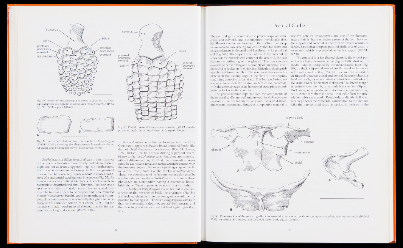
Fig. 53. Forefin of Brachypterygius extremus (BMNH R3177, holo-
type), depicted as a right fin in dorsal view (from McGowan [1997:
fig. 2B]). Scale equals 100 mm.
A
Fig. 54. Individual phalanx from the forefin of Platypterygius
(BMNH 47235), showing the characteristic house-brick shape.
A) planar and B) marginal views. Scale equals 40 mm.
Ophthalmosaurus differs from Ichthyosaurus in that most
of the forefin elements are not closely packed, so discrete
digits are not as readily apparent (Fig. 51). Furthermore,
the fin elements are rounded, except for the most proximal
ones, and all have coarsely rugose articular surfaces, indicative
of a substantial cartilaginous investment (Fig. 52). As
there are no clearly defined joint facets, it is not possible to
rearticulate disarticulated fins. Therefore, because most
specimens are disarticulated, there are few associated fore-
fins. The forefins appear to be broader and more rounded
than in Ichthyosaurus, but this could be an artifact of incomplete
data. For example, it was initially thought that Platypterygius
has a rounded forefin (McGowan, 1972C), but the
discovery of additional material showed that the fin was
remarkably long and slender (Wade, 1984).
Fig. 55. Partial forefin of Platypterygius australis (QM F3348), depicted
as a right fin in dorsal view. Scale equals 100 mm.
Brachypterygius, now known to range into the Early
Cretaceous, appears to have a broad, rounded forefin like
that of Ophthalmosaurus (Boulenger, 1904; McGowan,
1997). Indeed,-the fin bears a striking superficial resemblance
to that of Ophthalmosaurus, but there are some significant
differences (Fig. 53). First, the intermedium separates
the radius and ulna, and makes articular contact with
the humerus. Second, the rows of phalanges appear to lie
in vertical rows, more like the forefin of Ichthyosaurus.
Third, the elements tend to become_ rectangular distally,
not discoidal as they do in Ophthalmosaurus. Some of these
phalanges are rectangular, having a distinctive house-
brick shape. There appears to be upward of six digits.
The forefin of Platypterygius resembles that of Brachypterygius
in the presence of brick-like phalanges (Fig. 54),
and isolated elements from the two genera would be impossible
to distinguish. However, Platypterygius differs in
that the intermedium does not contact the humerus, and
the fin is long and slender, with at least eight digits (Fig.
55).
Pectoral Girdle
The pectoral girdle comprises the paired scapulae, coracoids,
and clavicles, and the unpaired interclavicle (Fig.
56). The coracoids come together in the midline, their deep
intercoracoidal facets being angled such that the distal end
of each element is elevated and lies dorsal to its proximal
end (Fig. 57E). The scapula articulates with the coracoid by
a facet on the anterolateral comer of the coracoid, the two
elements contributing to the glenoid. The clavicles are
joined together in a long and seemingly overlapping, inter-
digitating articulation in which it is difficult to distinguish
one element from the other. The conjoined clavicles articulate
with the leading edge of the shaft of the scapula,
continuing almost to its distal end. The T-shaped interclavicle
articulates with the ventral surface of the coracoids,
with the anterior edge of its horizontal cross-piece in intimate
contact with the clavicles.
The precise relationships between the components of
the pectoral girdle are well understood for Ophthalmosaurus
due to the availability of very well preserved three-
dimensional specimens. However, comparable material is
not available for Ichthyosaurus, and one of the shortcomings
of this is that the precise nature of the joint between
the scapula and coracoid is unclear. The present account is
largely based on a complete pectoral girdle of Ichthyosaurus
communis, which is preserved in ventral aspect (BMNH
R288).
The coracoid is a fan-shaped element, the widest part
of the fan being its medial edge (Fig. 57A-B). Most of this
medial edge is occupied by the intercoracoid facet (Fig.
57C), a thick, ellipsoidal area whose flattened surface is set
off from the vertical (Fig. 57A, E). This facet can be used to
distinguish between dorsal and ventral, because when it is
held vertically, as when paired coracoids are articulated,
the distal end of the element is elevated. The lateral margin
is entirely occupied by a second, but smaller, elliptical
thickening, which is divided into two unequal areas (Fig.
57D). Anteriorly, there is a small triangular facet for articulation
with the scapula. Posteriorly, a slightly hollowed
facet represents the coracoidal contribution to the glenoid.
Like the intercoracoid facet, its surface is inclined to the
Fig. 56. Reconstruction of the pectoral girdle of an essentially undistorted, acid-prepared specimen of Ichthyosaurus communis (BMNH
R288). A) ventral, B) anterior, and C) lateral views. Scale equals 100 mm.