shallow
depression
Fig. 39. Basisphenoid, dorsal view. A) Ichthyosaurus (OUMJ2242b). B) Ophthalmosaurus (LM 100’1949/49). C) Brachypterygius (SMC
J68516). D) unidentified ichthyosaur from the Upper Greensand (SMC B57962). 'Scale equals 40 mm.
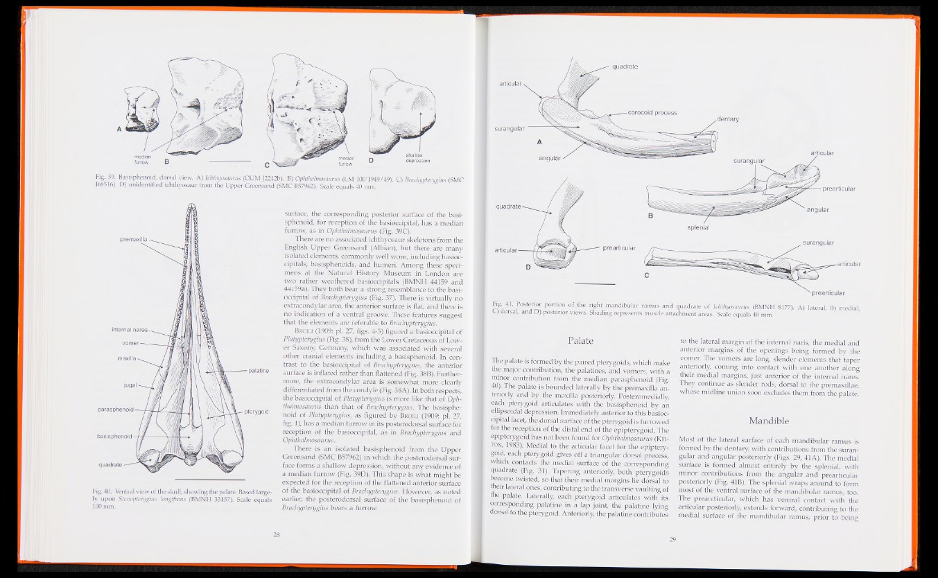
Fig. 40. Ventral view of the skull, showing the palate. Based largely
upon Stenopterygius longifrons (BMNH 33157). Scale equals
100 mm.
surface, the corresponding posterior surface of the basisphenoid,
for reception of the basioccipital, has a median
furrow, as in Ophthalmosaurus (Fig. 39C).
There are no associated ichthyosaur skeletons from the
- English Upper Greensand (Albian), but there are many
isolated elements, commonly welTvvom, including basioo-
cipitals, basisphenoids, and humeri. Among these specimens
at the Natural History Museum in London are
two rather weathered basioccipitals (BM.\) 1 44159 and
44159a). They both bear a strong resemblance to the basioccipital
of Brachypterygius (Fig, 37). .There is virtually no
extracondylar-area, the anterior surface is flat, and there is
no indication of a ventral groove. -These features suggest
that the elements are referable to Brachypterygius.
Broili (1909: plr27, figs. 4-5) figured a basioccipital of
Platypterygius (Fig. 38), from the Lower Cretaceous of Lower
Saxony, Germany, which was associated with several
other cranial elements including a basisphenoid. In contrast
to the basioccipital of Brachypterygius, the anterior
surface is inflated rather than flattened (Fig. 38B). Furthermore,
the extracondylar area is somewhat more clearly
-differentiated from the condyle (Fig. 38A). In both respects,
the basioccipital of Platypterygius is morelike that of Oph
thalmosaurus than that of Brachypterygius. The basisphenoid
of Platypterygius, as figured by Broil® (1909: pi. 27,
fig. 1), has amedian furrow in its posterodorsal surface for
reception of the basioccipital, as in Brachypterygius and
Qphthalrnostfurus,
There is an isolated basisphenoid from the-Upper
Greensand (SMC B579fi2) in which the posterodorsal surface
forms a shallow depression, without any evidence of
a median furrow (Fig. 39D). This shape is what might be
expected for the reception of the flattened anterior surface
of the basioccipital of Brachypterygius. However, as noted
earlier, the posterodorsal surface of the basisphenoid of
Brachypterygius beats a furrow.
quadrate
articular s
41- Posterior portion of the right mandibular ramus and quadrate of Ichthyosaurus (BMNH 8177). A) lateral, B) medial
G) dorsal, and D) posterior views. Shading represents muscle attachment areas. Scale equals 40 mm.
Palate
The palate is formed by the paired pterygoids, which make
the major contribution, the palatines, and vomers, with a
minor contribution from the median parasphenoid (Fig.
40). The palate is bounded laterally by the premaxilla anteriorly
and by the maxilla posteriorly. Posteromedially,
each pterygoid articulates with the basisphenoid by an
ellipsoidal depression. Immediately anterior to this basioccipital
facet, the dorsal surface of the pterygoid is furrowed
for the reception of the distal end of the epipterygoid. The
epipterygoid has not.been found for Ophthalmosaurus (Kir-
ton, 1983):. Medial to the articular facet for the epipterygoid,
each pterygoid gives off a triangular dorsal process,
which contacts the medial surface of the corresponding
quadrate (Fig. 31). Tapering anteriorly, both pterygoids
become twisted, so that their medial margins lie dorsal to
their lateral ones, contributing to the transverse vaulting of
the palate. Laterally, each pterygoid articulates with its
corresponding palatine in a lap joint, the palatine lying
dorsal to the pterygoid. Anteriorly, the palatine contributes
to the lateral margin of the internal naris, the medial and
anterior margins of the openings being formed by the
vomer. The vomers are long, slender elements that taper
anteriorly, coming into contact with one another along
their medial margins, just anterior of the internal nares.
They continue as slender rods, dorsal to the premaxillae,
Whose midline union soon excludes them from the palate.
Mandible
Most of the lateral surface of each mandibular ramus is
formed by the dentary, with contributions from the suran-
gular and angular posteriorly (Figs. 29, 41A). The medial
surface is formed almost entirely by the splenial, with
minor contributions from the angular and prearticular
posteriorly (Fig. 41B). The splenial wraps around to form
most of the ventral surface of the mandibular ramus, too.
The prearcticular, which has ventral contact with the
articular posteriorly, extends forward, contributing to the
medial surface of the mandibular ramus, prior to being