dorsal process (?) anterior ventral process (?)
facet for fibula for tibia (?)
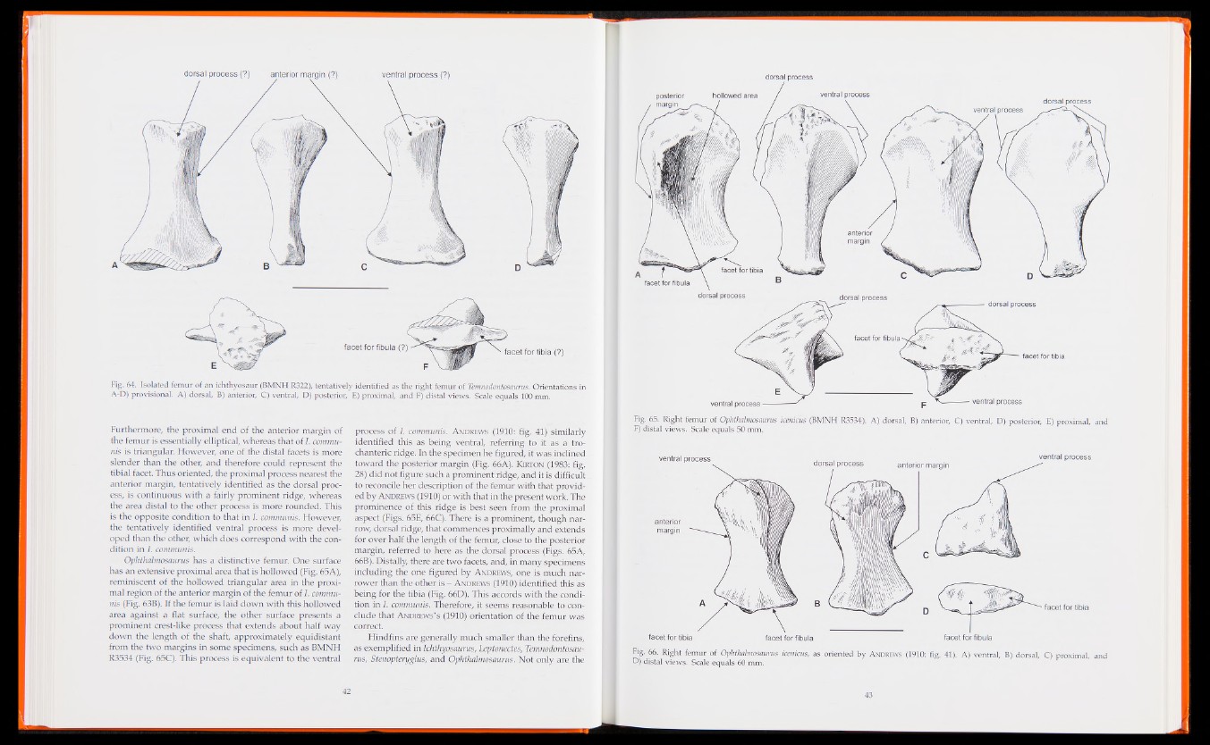
Fig. 64. Isolated femur of an ichthyosaur (BMNH R322), tentatively identified as the right femur of Temnodontosaurus. Orientations in
A-D) provisional. A) dorsal, B) anterior, C) ventral, D) posterior, E) proximal, and F) distal views. Scale equals 100 mm.
Furthermore, the proximal end of the anterior margin of
the femur is essentially elliptical, whereas that of I. communis
is triangular. However, one of the distal facets is more
slender than the other, and therefore could represent the
tibial facet. Thus oriented, the proximal process nearest the
anterior margin, tentatively identified as the dorsal process,
is continuous with a fairly prominent ridge, whereas
the area distal to the other process is more rounded. This
is the opposite condition to that in I. communis. However,
the tentatively identified ventral process is more developed
than the other, which does correspond with the condition
in 1. communis.
Ophthalmosaurus has a distinctive femur. One surface
has an extensive proximal area that is hollowed (Fig. 65A),
reminiscent of the hollowed triangular area in the proximal
region of the anterior margin of the femur of I. communis
(Fig. 63B). If the femur is laid down with this hollowed
area against a flat surface, the other surface presents a
prominent crest-like process that extends about half way
down the length of the shaft, approximately equidistant
from the two margins in some specimens, such as BMNH
R3534 (Fig. 65C). This process is equivalent to the ventral
process of 1. communis. Andrews (1910: fig. 41) similarly
identified this as being ventral, referring to it as a trochanteric
ridge. In the specimen he figured, it was inclined
toward the posterior margin (Fig. 66A). Kirton (1983: fig.
28) did not figure such a prominent ridge, and it is difficult
to reconcile her description of the femur with that provided
by A ndrews (1910) or with that in the present work. The
prominence of this ridge is best seen from the proximal
aspect (Figs. 65E, 66C). There is a prominent, though narrow,
dorsal ridge, that commences proximally and extends
for over half the length of the femur, close to the posterior
margin, referred to here as the dorsal process (Figs. 65A,
66B). Distally, there are two facets, and, in many specimens
including the one figured by A ndrews, one is much narrower
than the other is - A ndrews (1910) identified this as
being for the tibia (Fig. 66D). This accords with the condition
in I. communis. Therefore, it seems reasonable to conclude
that Andrews’s (1910) orientation of the femur was
correct.
Hindfins are generally much smaller than the forefins,
as exemplified in Ichthyosaurus, Leptonectes, Temnodontosaurus,
Stenopterygius, and Ophthalmosaurus. Not only are the
dorsal process
dorsal process
dorsal process
ventral process
facet for tibia
A) dorsal, B) anterior, C) ventral, D) posterior, E) proximal, and
ventral process^
Fig. 65. Right femur of Ophthalmosaurus icenicus (BMNH R3534).
F) distal views. Scale equals 50 mm.
Fig. 66. Right femur of Ophthalmosaurus icenicus, as oriented b y A ndrews (1910: fig. 41). A ) ventral, B) dorsal, C) proximal, and
D) distal views. Scale equals 60 mm.