teeth, including those few specimens which have been
preserved with the jaws agape. This is presumably because
the teeth are so firmly attached.
As seen in serial sections, the dental groove does not
extend the entire depth of the snout or mandible (Fig. 42).
Instead, it is delimited by an essentially horizontal shelf,
extending from the premaxilla, or from the dentary, against
which the base of the teeth abut. The teeth typically have
much of the root exposed, giving the impression that some
displacement from the groove has occurred. However, this
is unlikely, as revealed by a three-dimensional rostral segment
from a large skull that is probably referable to Temn-
odontosaurus platyodon (Fig. 45). Several of the teeth are
exposed in their entirety, and their roots extend well beyond
the distal edge of the dental groove. However, the
proximal ends of the roots are in contact with the horizontal
shelf, showing that they have not been displaced.
Hyoid
The hyoid apparatus, which is seldom exposed, appears to
comprise a pair of stout rods that lie medial to the mandibular
rami, at about the level of the orbits (Fig. 43). Sollas
(1916: fig. 15) described and figured a fairly complex apparatus,
which he admitted did not resemble that of any
extant reptile. KlRTON (1983) believed the reason for this
seeming complexity is that some displaced ribs were associated
with the hyoid apparatus in Sollas’s specimen. This
seems more than likely, and can also be seen elsewhere.
Thus, in BMNH 49203 (I. communis), a rod-like element lies
in contact with the right ramus of the hyoid (not shown in
Fig. 43), and this could be interpreted as part of the apparatus.
However, one end of this bone has a shallow furrow,
with a slightly raised edge, which is identified as a displaced
rib. The hyoid is exposed on one side in BMNH
R1158, a mature specimen of T. platyodon, and is exactly
similar to that of I communis.
Forefin
Humerus
Ichthyosaurus has a compact humerus, with a short shaft.
The proximal end is robust, whereas the distal end is much
thinner, and is elongated in the plane of the rest of the
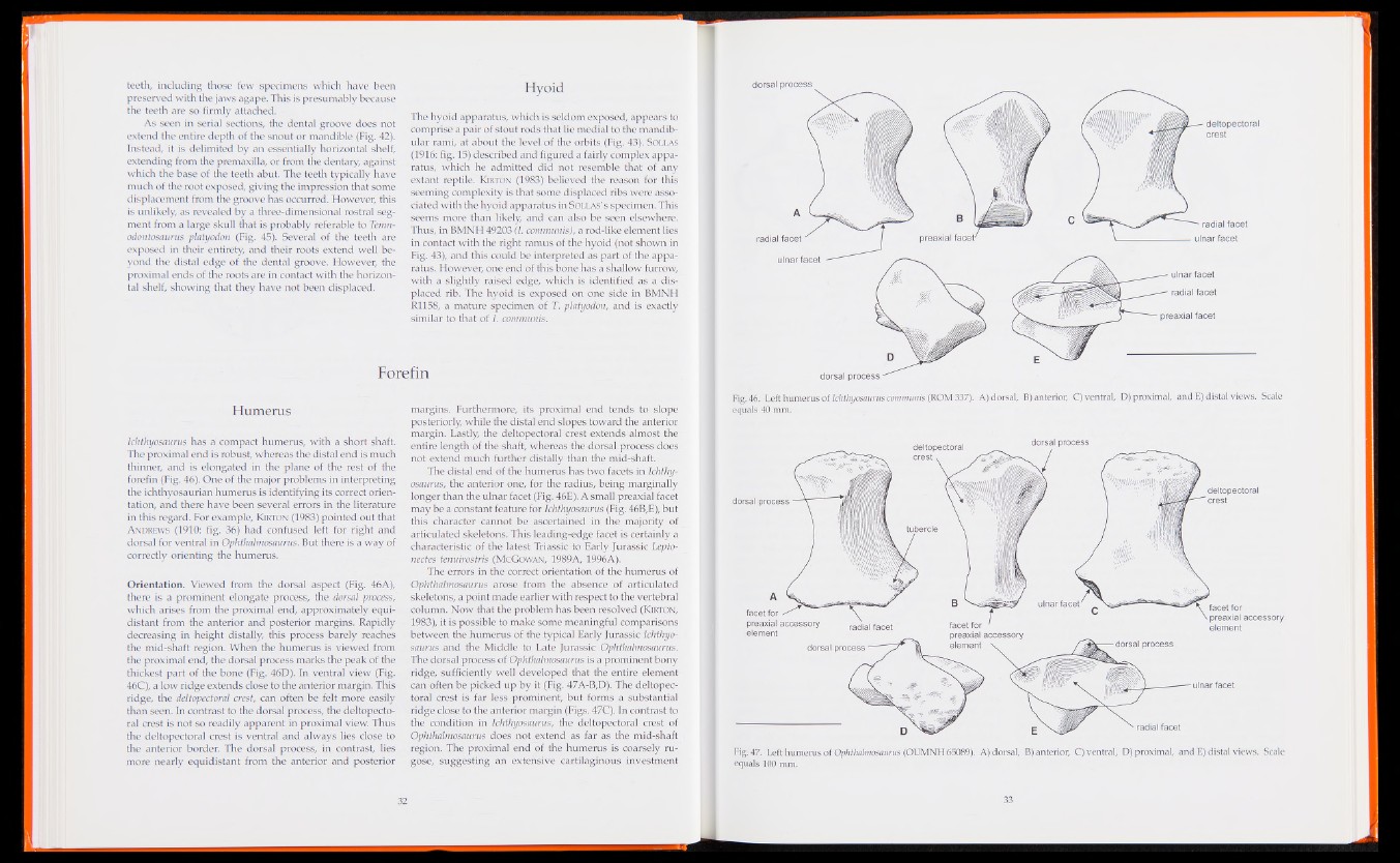
forefin (Fig. 46). One of the major problems in interpreting
the ichthyosaurian humerus is identifying its correct orientation,
and there have been several errors in the literature
in this regard. For example, Kjrton (1983) pointed out that
Andrews (1910: fig. 36) had confused left for right and
dorsal for ventral in Ophthalmosaurus. But there is a way of
correctly orienting the humerus.
Orientation. Viewed from the dorsal aspect (Fig. 46A),
there is a prominent elongate process, the dorsal process,
which arises from the proximal end, approximately equidistant
from the anterior and posterior margins. Rapidly
decreasing in height distally, this process barely reaches
the mid-shaft region. When the humerus is viewed from
the proximal end, the dorsal process marks the peak of the
thickest part of the bone (Fig. 46D). In ventral view (Fig.
46C), a low ridge extends close to the anterior margin. This
ridge, the deltopectoral crest, can often be felt more easily
than seen. In contrast to the dorsal process, the deltopectoral
crest is not so readily apparent in proximal view. Thus
the deltopectoral crest is ventral and always lies close to
the anterior border. The dorsal process, in contrast, lies
more nearly equidistant from the anterior and posterior
margins. Furthermore, its proximal end tends to slope
posteriorly, while the distal end slopes toward the anterior
margin. Lastly, the deltopectoral crest extends almost the
entire length of the shaft, whereas the dorsal process does
not extend much further distally than the mid-shaft.
The distal end of the humerus has two facets in Ichthyosaurus,
the anterior one, for the radius, being marginally
longer than the ulnar facet (Fig. 46E). A small preaxial facet
may be a constant feature for Ichthyosaurus (Fig. 46B,E), but
this character cannot be ascertained in the majority of
articulated skeletons. This leading-edge facet is certainly a
characteristic of the latest Triassic to Early Jurassic Lepto-
nectes tenuirostris (McGowan, 1989A, 1996A).
The errors in the correct orientation of the humerus of
Ophthalmosaurus arose from the absence of articulated
skeletons, a point made earlier with respect to the vertebral
column. Now that the problem has been resolved (Kirton,
1983), it is possible to make some meaningful comparisons
between the humerus of the typical Early Jurassic Ichthyosaurus
and the Middle to Late Jurassic Ophthalmosaurus.
The dorsal process of Ophthalmosaurus is a prominent bony
ridge, sufficiently well developed that the entire element
can often be picked up by it (Fig. 47A-B,D). The deltopectoral
crest is far less prominent, but forms a substantial
ridge close to the anterior margin (Figs. 47C). In contrast to
the condition in Ichthyosaurus, the deltopectoral crest of
Ophthalmosaurus does not extend as far as the mid-shaft
region. The proximal end of the humerus is coarsely rugose,
suggesting an extensive cartilaginous investment
dorsal process
deltopectoral
crest
—■ radial facet
radial facet preaxial facet' ulnar facet
ulnar facet
radial facet
preaxial facet
dorsal process -—
Fig. 46. Left humerus of Ichthyosaurus communis (ROM 337). A) dorsal, B) anterior, C) ventral, D) proximal, and E) distal views. Scale
equals 40 mm.
deltopectoral dorsal process
crest v
deltopectoral
dorsal process crest
tubercle
ulnar facet' facet for
preaxial accessory
element
facet for / ^
preaxial accessory
element
facet for '
preaxial accessory
element v
radial facet
dorsal process dorsal process
ulnar facet
radial facet
Fig. 47. Left humerus of Ophthalmosaurus (OUMNH 65089). A) dorsal, B) anterior, C) ventral, D) proximal, and E) distal views. Scale
equals 100 mm.