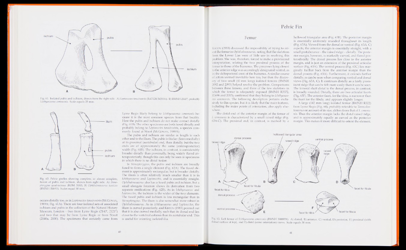
ischium
ischium
Fig. 61. Isolated pubis and ischium, drawn from the right side. A) Leptonectes tenuirostris (BATGM M3556). B) BMNH 2140*, probably
Ichthyosaurus communis. Scale equals 25 mm.
Fig. 62. Pelvic girdles showing complete, or almost complete,
fusion of pubis and ischium, shown from right side. A) Stenopterygius
quadrisdssus (ROM 3180). B) Ophthalmosaurus icenicus
(BMNH R4693). Scales equal 50 mm.
occurs distally too, as in Leptonectes tenuirostris (McGowan,
1989A; Fig. 61 A). There are four isolated sets of associated
ischium and pubis in the collection of the Natural History
Museum, London - two from Lyme Regis (2140*, 2121*)
and two that may be from Lyme Regis or from Street
(2140a, 2100). The specimens that certainly came from
Lyme Regis likely belong to Ichthyosaurus communis because
it is the most common species from that locality.
Here the pubis and ischium do not make contact distally
(Fig. 61B). The other specimens are both fused distally and
probably belong to Leptonectes tenuirostris, a species commonly
found at Street (McGowan, 1989A).
The pubis and ischium are similar in length to each
other and to the ilium. The pubis is thicker (lateromedially)
at its proximal (acetabular) end, than distally, but the two
ends are of approximately the same (anteroposterior)
width (Fig. 61B). The ischium, in contrast, is considerably
broader distally than proximally, being widely flared an-
teroposteriorly, though this can only be seen in specimens
in which there is no distal fusion.
In Stenopterygius, the pubis and ischium are broadly
fused to form a single element (Fig. 62A). The fused element
is approximately rectangular, but is broader distally.
The ilium is often relatively much smaller than it is in
Ichthyosaurus and Leptonectes, and' is essentially straight.
Ophthalmosaurus also has a fused pubis and ischium, but a
small elongate foramen shows its derivation from two
separate ossifications (Fig. 62B). As in Ichthyosaurus and
Leptonectes, the ischium is the wider of the two elements.
The fused pubis and ischium is less rectangular than in
Stenopterygius. The ilium is also somewhat more robust in
Ophthalmosaurus. As in Ichthyosaurus and Leptonectes, the
ilium is curved posteriorly, and K erton (1983) pointed out
that it is also curved medially, such that its dorsal end lies
closer to the vertebral column than its acetabular end. This
is useful for orienting isolated ilia.
Pelvic Fin
Femur
Klrton (1983) discussed the impossibility of trying to orient
the femur in Ophthalmosaurus, noting that the skeletons
from the Lower Lias were of little use in resolving this
problem. She was, therefore, forced to make a provisional
interpretation, relating the two proximal process of the
femur to those of the humerus. The processes lying closest
to the anterior edge was accordingly designated ventral, as
in the deltopectoral crest of the humerus. A similar course
of action seemed inevitable here too, but then the discovery
of two small (41 mm long) isolated femora (BMNH
2092 and 2093) helped resolve the problem. Comparisons
between these femora, and three of the few skeletons in
which the femur is adequately exposed (BMNH R3372,
R1696 and 2013), confirmed that they belong to Ichthyosaurus
communis. The following description pertains exclusively
to this species, but it is likely that the main features,
including the major points of orientation, also apply elsewhere.
The distal end of the anterior margin of the femur of
I communis is characterized by a small raised ridge (Fig.
63A-C). The proximal end,' in contrast, is marked by a
hollowed triangular area (Fig. 63B). The posterior margin
is essentially uniformly rounded throughout its length
(Fig. 63A). Viewed from the dorsal or ventral (Fig. 63A, C)
aspects, the anterior margin is essentially straight, with a
small protuberance - the raised ridge - distally. The posterior
margin, however, is markedly curved, and flared pos-
terodistally. The dorsal process lies close to the anterior
margin, and is just an extension of the proximal articular
surface (Fig. 63A). The ventral process (Fig. 63C) lies marginally
further back from the anterior margin than the
dorsal process (Fig. 63A). Furthermore, it extends further
distally, as can be seen when comparing ventral and dorsal
views (Fig. 63A, C). It continues distally as a fairly prominent
ridge that can be felt more easily than it can be seen.
The femoral shaft distal to the dorsal process, in contrast,
is broadly rounded. Distally, there are two articular facets
(Fig. 63F). The anterior facet, for the tibia, is narrower than
the facet for the fibula, and appears slightly longer.
A large (142 mm long) isolated femur (BMNH R322)
from Lyme Regis (Fig. 64), probably referable to Temnodon-
tosaurus on account of its size, differs from that of I. communis.
Thus the anterior margin lacks the distal raised ridge,
and is approximately equally as curved as the posterior
margin. This makes it more difficult to orient the element.
dorsal process
raised ridge
hollowed triangular area
raised ridge ventral process
facet for tibia
dorsal process
Fig. 63. Left femur of Ichthyosaurus communis (BMNH 300093X). A) dorsal, B) anterior, C) ventral, D) posterior, E) proximal (with
dorsal surface at top), and F) distal (same orientation) views. Scale equals 30 mm.