facets
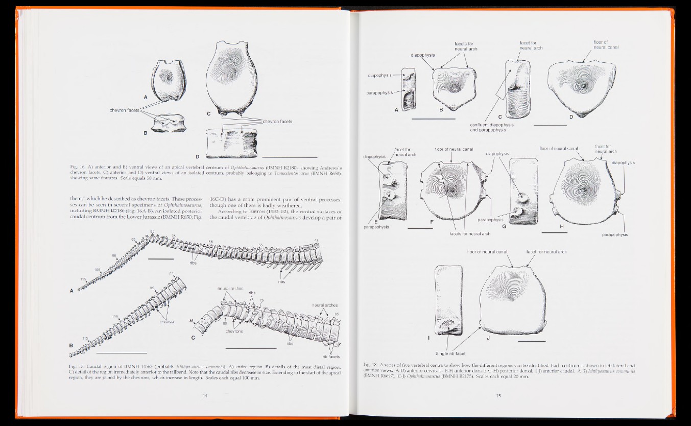
Fig. 16. A) anterior and B) ventral views of an apical vertebral centrum of Ophthalmosaurus (BMNH R2180), showing Andrews’s
chevron facets. C) anterior and D) ventral views of an isolated centrum, probably belonging to Temnodontosaurus (BMNH R650),
showing same features. Scale equals 50 mm.
them,” which he described as chevron facets. These processes
can be seen in several specimens of Ophthalmosaurus,
including BMNH R2180 (Fig. 16A-B). An isolated posterior
caudal centrum from the Lower Jurassic (BMNH R650; Fig.
16C-D) has a more prominent pair of ventral processes,
though one of them is badly weathered.
Accordin g to Kirton (1983: 82), the ventral surfaces of
the caudal vertebrae of Ophthalmosaurus develop a pa ir of
Fig. 17. Caudal region of BMNH 14563 (probably Ichthyosaurus communis). A) entire region. B) details of the most distal region.
C) detail of the region immediately anterior to the tailbend. Note that the caudal ribs decrease in size. Extending to the start of the apical
region, they are joined by the chevrons, which increase in length. Scales each equal 100 mm.
facets for
neural arch
diapophysis
parapophysis
facet for
neural arch
confluent diapophysis
and parapophysis
facet for
parapophysis
parapophysis
Single rib facet
diapophysis
floor of neural canal fl°or of neural canal
diapophysis
5 f l
facets for neural arch
floor of neural canal facet for neural arch
Fig. 18. A series of five vertebral centra to show how the different regions can be identified. Each centrum is shown in left lateral and
anterior views. A-D) anterior cervicals; E-F) anterior dorsal; G-H) posterior dorsal; I-J) anterior caudal. A-B) Ichthyosaurus communis
(BMNH R6697); C-J) Ophthalmosaurus (BMNH R2175). Scales each equal 20 mm.