condyle exoccipital facets
basioccipital peg
median ridge
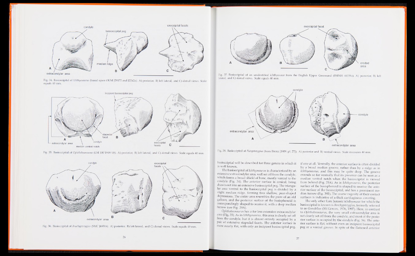
Fig. 34. Basiocdpital of Ichthyosaurus (based upon OUM J29172 and J2242a). A) posterior, B) left lateral, and C) dorsal views. Scale
equals 40 mm.
exoccipital
facets C
incipient basioccipital peg
A
— extracondylar area condyle
ventral notch
Fig. 35. Basiocdpital of Ophthalmosaurus (LM 100 1949/48). A) posterior, B) left lateral, and C) dorsal views. Scale equals 40 mm.
condyle exoccipital
B
extracondylar area
facets
Fig. 36. Basiocdpital of Brachypterygius (SMC J68516). A) posterior, B) left lateral, and C) dorsal views. Scale equals 40 mm.
exoccipital facet
eroded
area
Fig. 37. Basioccipital of an unidentified ichthyosaur from the English Upper Greensand (BMNH 44159a). A) posterior, B) left
lateral, and C) dorsal views. Scale equals 40 mm.
condyle
extracondylar area
A
condyle
extracondylar area
Fig. 38. Basioccipital of Platypterygius (from Broili [1909: pi. 27]). A) posterior and B) ventral views. Scale measures 40 mm.
basiocdpital will be described for three genera in which it
is well kno\vn.
The basioccipital of Ichthyosaurus is characterized by an
extensive extracondylar area, well set off from the condyle,
which forms a broad shield of bone, mostly ventral to the
condyle (Fig. 34). The anterior surface is conical, being
drawn out into an extensive basioccipital peg. The triangular
area ventral to the basioccipital peg is divided by a
slight median ridge, forming two shallow, pear-shaped
depressions. The entire area resembles the prow of an old
galleon, and the posterior surface of the basisphenoid is
correspondingly shaped to receive it, with a deep median
furrow (see Fig. 39A),
Ophthalmosaurus has a far less extensive extracondylar
area (Fig. 35), As in Ichthyosaurus, this area is clearly set off
from the condyle, but it is almost entirely occupied by a
pair of extensive stapedial facets. The anterior surface is
more nearly flat, with only an incipient basioccipital peg,
if one at all. Ventrally, the anterior surface is often divided
by a broad median groove, rather than by a ridge as in
Ichthyosaurus, and this may be quite deep. The groove
extends so far ventrally that its presence can be seen as a
median ventral notch when the basioccipital is viewed
from behind (Fig. 35A). As in Ichthyosaurus, the posterior
surface of the basisphenoid is shaped to receive the anterior
surface of the basioccipital, and has a prominent median
furrow (Fig. 39B). The coarse rugosity of their contact
surfaces is indicative of a thick cartilaginous covering.
The only other Late Jurassic ichthyosaur for which the
basioccipital is known is Brachypterygius, formerly referred
to as GremieMus (McGowan*, 1976, 1997). Here, in contrast
to Ophthalmosaurus, the very small extracondylar area is
not clearly set off from the condyle, and most of the posterior
surface is occupied by the condyle (Fig. 36). The anterior
surface is flat, without even an incipient basioccipital
peg or a ventral groove. In spite of the Battened anterior