facet for neural arch
Fig. 2. A) an anterior vertebra, where the diapophysis contacts the facet for the neural arch. B) a more posterior vertebra where contact
between the two has been lost. Both in left lateral view. Scale equals 50 mm.
bly all referable to Ichthyosaurus communis. This is because
many determinate specimens belong to this common species,
and the indeterminate ones are entirely consistent
with them. What makes this material from nodules so
important is that the individual bones are virtually undistorted,
and their precise spatial relationships are known.
Ichthyosaurian vertebrae vary in form throughout the
vertebral column. It is thus possible to identify an isolated
vertebral centrum with a given region. However, as Kirton
(1983) has pointed out, there are no sharply defined boundaries
between one region and the next, in spite of the attempts
of some authorities to define them. A ppleby (1956),
for example, took the boundary between cervical and dorsal
vertebrae in Ophthalmosaurus as the point where the
diapophysis (the most dorsal rib facet) loses contact with
the facet for the neural arch (Fig. 2). However, the point at
which this occurs, which is somewhat variable, lays posterior
to the position of the pectoral girdle. Indeed, the cervical
vertebrae merge with the dorsal vertebrae without
interruption, as do the ribs that articulate with them.
Therefore, it is meaningless to attempt a morphological
distinction between the two, and these respective regions
dorsal
intercentrum
axial centrum
parapophysis
facet for atlantal
axis
D
facets
for
neural arches
atlantal
concavity
facet for atlantal
intercentrum
axial
concavity
axial
intercentrum
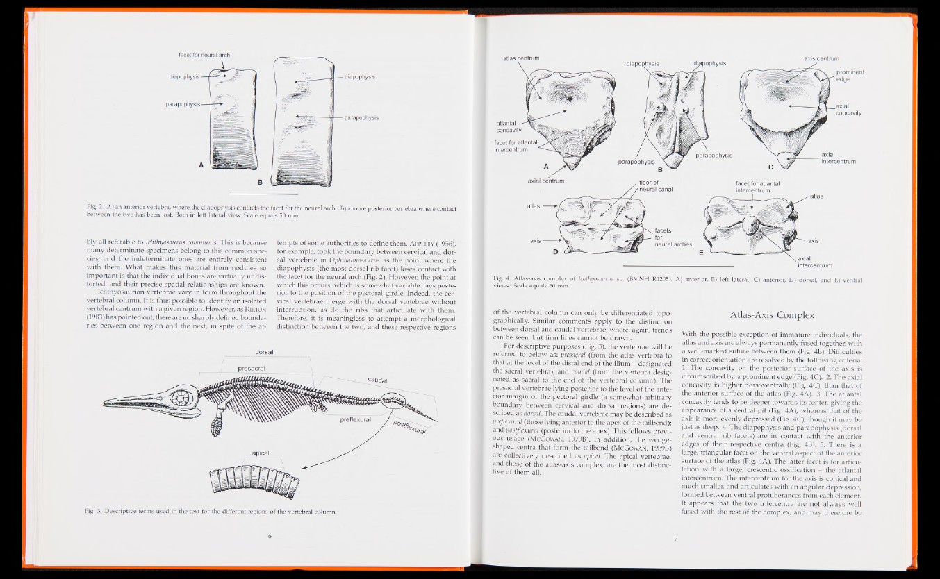
Fig. 4. Atlas-axis complex of Ichthyosaurus sp. (BMNH R1205). A) anterior, B) left lateral, C) anterior, D) dorsal, and E) ventral
views. Seale equals SO mm.
of the vertebral column can only be differentiated topographically.
Similar comments apply to the distinction
between dorsal and caudal vertebrae, where, again, trends
can be seen, but firm lines cannot be drawn.
For descriptive purposes (Fig. 3), the vertebrae will be
referred to below as: ■ presacral (from the atlas vertebra to
that at the level of the distal end of the ilium - designated
the sacral vertebra); and caudal (from the vertebra designated
as sacral to the end of the vertebral column). The
presacral vertebrae lying posterior to the level of the anterior
margin of the pectoral girdle (a somewhat arbitrary
boundary between cervical and dorsal regions) are described
as dorsal. The caudal vertebrae may be described as
preflexural (those lying anterior to the apex of the tailbend);
and pustflexural (posterior to the apex). This follows previous
usage (McGowan, 197911). In addition, the wedge-
shaped centra that form the tailbend (McGowan, 1989B)
are collectively described as apical. The apical vertebrae,
and those of the atlas-axis complex, are the most distinctive
of them all.
Atlas-Axis Complex
With the possible exception of immature individuals, the
atlas and axis are always permanently fused together, with
a well-marked suture between them (Fig. 4B). Difficulties
in correct orientation are resolved by the following criteria:
1. The concavity on the posterior surface of the axis is
circumscribed by a prominent edge (Fig. 4C). 2. The axial
concavity is higher dorsoventrally (Fig. 4C), than that of
the anterior surface of the atlas (Fig. 4A). 3. The atlantal
concavity tends to be deeper towards its center, giving the
appearance of a central pit (Fig. 4A), whereas (hat of the
axis is more evenly depressed (Fig. 4C), though it may be
just as deep. 4. The diapophysis and parapophysis (dorsal
and ventral rib facets) are in contact with the anterior
edges of their respective centra (Fig. 4B). 5. There is a
large, triangular facet on the ventral aspect of the anterior
surface of the atlas (Fig. 4A). The latter facet is for articulation
with a large, crescentic ossification - the atlantal
intercentrum. The intercentrum for the axis is conical and
much smaller, and articulates with an angular depression,
formed between ventral protuberances from each element.
If appears that the two intercentra are not always well
fused with the rest of the complex, and may therefore be