lachrymal
parietal
supratemporal
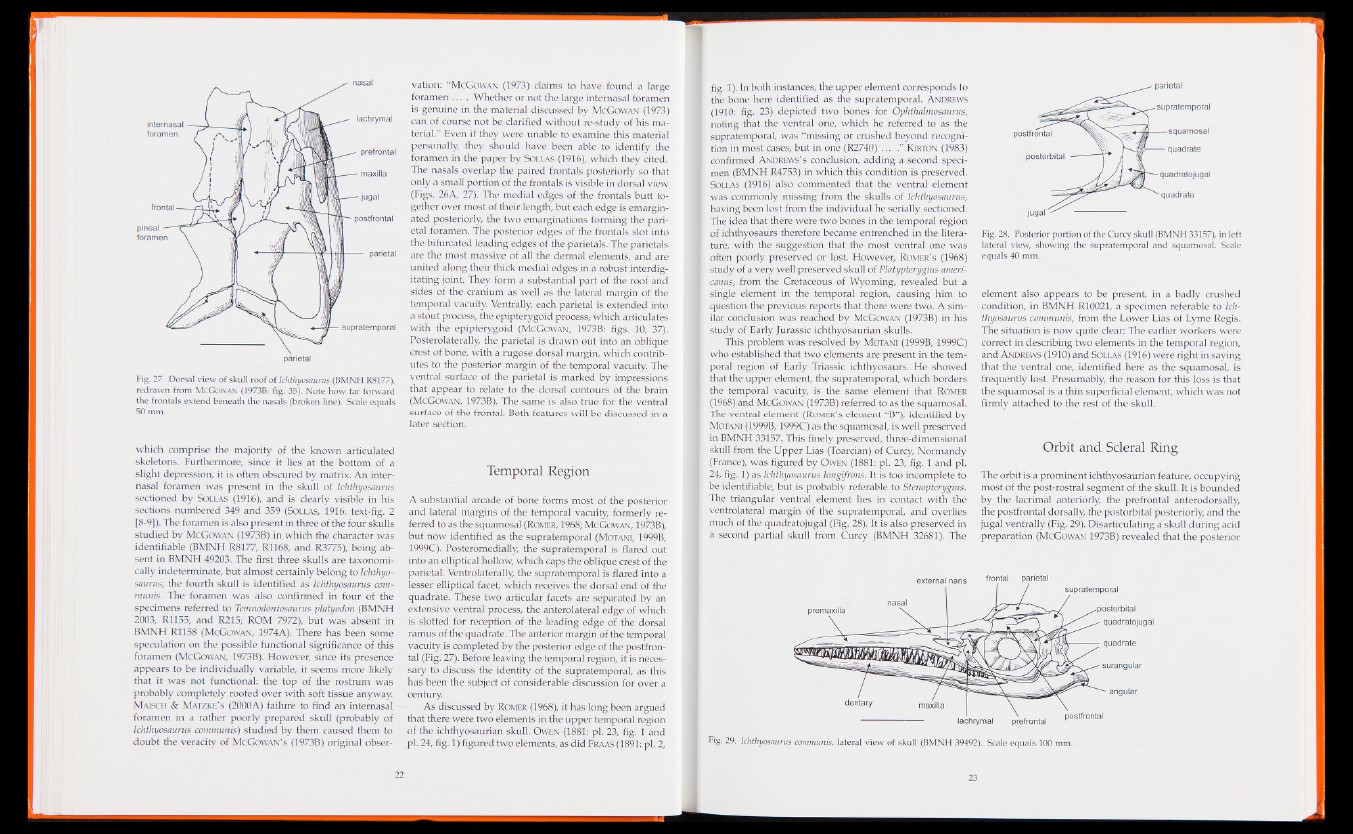
Fig. 27. Dorsal view of skull roof of Ichthyosaurus (BMNH R8177),
redrawn from McGowan (1973B: fig. 35). Note how far forward
the frontals extend beneath the nasals (broken line). Scale equals
50 mm.
vation: “McGowan (1973) claims to have found a large
foramen . . . . Whether or not the large intemasal foramen
is genuine in the material discussed by McGowan (1973)
can of course not be clarified without re-study of his material.”
Even if they were unable to examine this material
personally they should have been able to identify the
foramen in the paper by Sollas (1916), which they cited.
The nasals overlap the paired frontals posteriorly so that
only a small portion of the frontals is visible in dorsal view
(Figs. 26A, 27). The medial edges of the frontals butt together
over most of their length, but each edge is emargin-
ated posteriorly, the two emarginations forming the parietal
foramen. The posterior edges of the frontals slot into
the bifurcated leading edges of the parietals. The parietals
are the most massive of all the dermal elements, and are
united along their thick medial edges in a robust interdig-
itating joint. They form a substantial part of the roof and
sides of the cranium as well as the lateral margin of the
temporal vacuity. Ventrally, each parietal is extended into
a stout process, the epipterygoid process, which articulates
with the epipterygoid (McGowan, 1973B: figs. 10, 37).
Posterolaterally, the parietal is drawn out into an oblique
crest of bone, with a rugose dorsal margin, which contributes
to the posterior margin of the temporal vacuity. The
ventral surface of the parietal is marked by impressions
that appear to relate to the dorsal contours of the brain
(McGowan, 1973B). The same is also true for the ventral
surface of the frontal. Both features will be discussed in a
later section.
which comprise the majority of the known articulated
skeletons. Furthermore, since it lies at the bottom of a
slight depression, it is often obscured by matrix. An internasal
foramen was present in the skull of Ichthyosaurus
sectioned by Sollas (1916), and is clearly visible in his
sections numbered 349 and 359 (Sollas, 1916: text-fig. 2
[8-9]). The foramen is also present in three of the four skulls
studied by McGowan (1973B) in which the character was
identifiable (BMNH R8177, R1168, and R3775), being absent
in BMNH 49203. The first three skulls are taxonomi-
cally indeterminate, but almost certainly belong to Ichthyosaurus;
the fourth skull is identified as Ichthyosaurus communis.
The foramen was also confirmed in four of the
specimens referred to Temnodontosaurus platyodon (BMNH
2003, R1155, and R215; ROM 7972), but was absent in
BMNH R1158 (McGowan, 1974A). There has been some
speculation on the possible functional significance of this
foramen (McGowan, 1973B). However, since its presence
appears to be individually variable, it seems more likely
that it was not functional: the top of the rostrum was
probably completely roofed over with soft tissue anyway.
Maisch & Matzke’s (2000A) failure to find an internasal
foramen in a rather poorly prepared skull (probably of
Ichthyosaurus communis) studied by them caused them to
doubt the veracity of McGowan’s (1973B) original obser-
Temporal Region
A substantial arcade of bone forms most of the posterior
and lateral margins of the temporal vacuity, formerly referred
to as the squamosal (RomER, 1968; McGowan, 1973B),
but now identified as the supratemporal (Motani, 1999B,
1999C). Posteromedially, the supratemporal is flared out
into an elliptical hollow, which caps the oblique crest of the
parietal. Ventrolaterally, the supratemporal is flared into a
lesser elliptical facet, which receives the dorsal end of the
quadrate. These two articular facets are separated by an
extensive ventral process, the anterolateral edge of which
is slotted for reception of the leading edge of the dorsal
ramus of the quadrate. The anterior margin of the temporal
vacuity is completed by the posterior edge of the postfrontal
(Fig. 27). Before leaving the temporal region, it is necessary
to discuss the identity of the supratemporal, as this
has been the subject of considerable discussion for over a
century.
As discussed by Romer (1968), it has long been argued
that there were two elements in the upper temporal region
of the ichthyosaurian skull. Owen (1881: pi. 23, fig. 1 and
pi. 24, fig. 1) figured two elements, as did Fraas (1891: pi. 2,
fig. 1). In both instances, the upper element corresponds to
the bone here identified as the supratemporal. Andrews
(1910: fig. 23) depicted two bones for Ophthalmosaurus,
noting that the ventral one, which he referred to as the
supratemporal, was “missing or crushed beyond recognition
in most cases, but in one (R2740) ... .” Kirton (1983)
confirmed Andrews’s conclusion, adding a second specimen
(BMNH R4753) in which this condition is preserved.
Sollas (1916) also commented that the ventral element
was commonly missing from the skulls of Ichthyosaurus,
having been lost from the individual he serially sectioned.
The idea that there were two bones in the temporal region
of ichthyosaurs therefore became entrenched in the literature,
with the suggestion that the most ventral one was
often poorly preserved or lost. However, Romer’s (1968)
study of a very well preserved skull of Platypterygius ameri-
canus, from the Cretaceous of Wyoming, revealed but a
single element in the temporal region, causing him to
question the previous reports that there were two. A similar
conclusion was reached by McGowan (1973B) in his
study of Early Jurassic ichthyosaurian skulls.
This problem was resolved by Motani (1999B, 1999C)
who established that two elements are present in the temporal
region of Early Triassic ichthyosaurs. He showed
that the upper element, the supratemporal, which borders
the temporal vacuity, is the same element that Romer
(1968) and McGowan (1973B) referred to as the squamosal.
The ventral element (Romer’s element “B”), identified by
Motani (1999B, 1999C) as the squamosal, is well preserved
in BMNH 33157. This finely preserved, three-dimensional
skull from the Upper Lias (Toarcian) of Curcy, Normandy
(France), was figured by Owen (1881: pi. 23, fig. 1 and pi.
24, fig. 1) as Ichthyosaurus longifrons. It is too incomplete to
be identifiable, but is probably referable to Stenopterygius.
The triangular ventral element lies in contact with the
ventrolateral margin of the supratemporal, and overlies
much of the quadratojugal (Fig. 28). It is also preserved in
a second partial skull from Curcy (BMNH 32681). The
Fig. 28. Posterior portion of the Curcy skull (BMNH 33157), in left
lateral view, showing the supratemporal and squamosal. Scale
equals 40 mm.
element also appears to be present, in a badly crushed
condition, in BMNH R10021, a specimen referable to Ichthyosaurus
communis, from the Lower Lias of Lyme Regis.
The situation is now quite clear: The earlier workers were
correct in describing two elements in the temporal region,
and Andrews (1910) and Sollas (1916) were right in saying
that the ventral one, identified here as the squamosal, is
frequently lost. Presumably, the reason for this loss is that
the squamosal is a thin superficial element, which was not
firmly attached to the rest of the skull.
Orbit and Scleral Ring
The orbit is a prominent ichthyosaurian feature, occupying
most of the post-rostral segment of the skull. It is bounded
by the lacrimal anteriorly, the prefrontal anterodorsally,
the postfrontal dorsally, the postorbital posteriorly, and the
jugal ventrally (Fig. 29). Disarticulating a skull during acid
preparation (McGowan 1973B) revealed that the posterior
Fig. 29. Ichthyosaurus communis, lateral view of skull (BMNH 39492). Scale equals 100 mm.