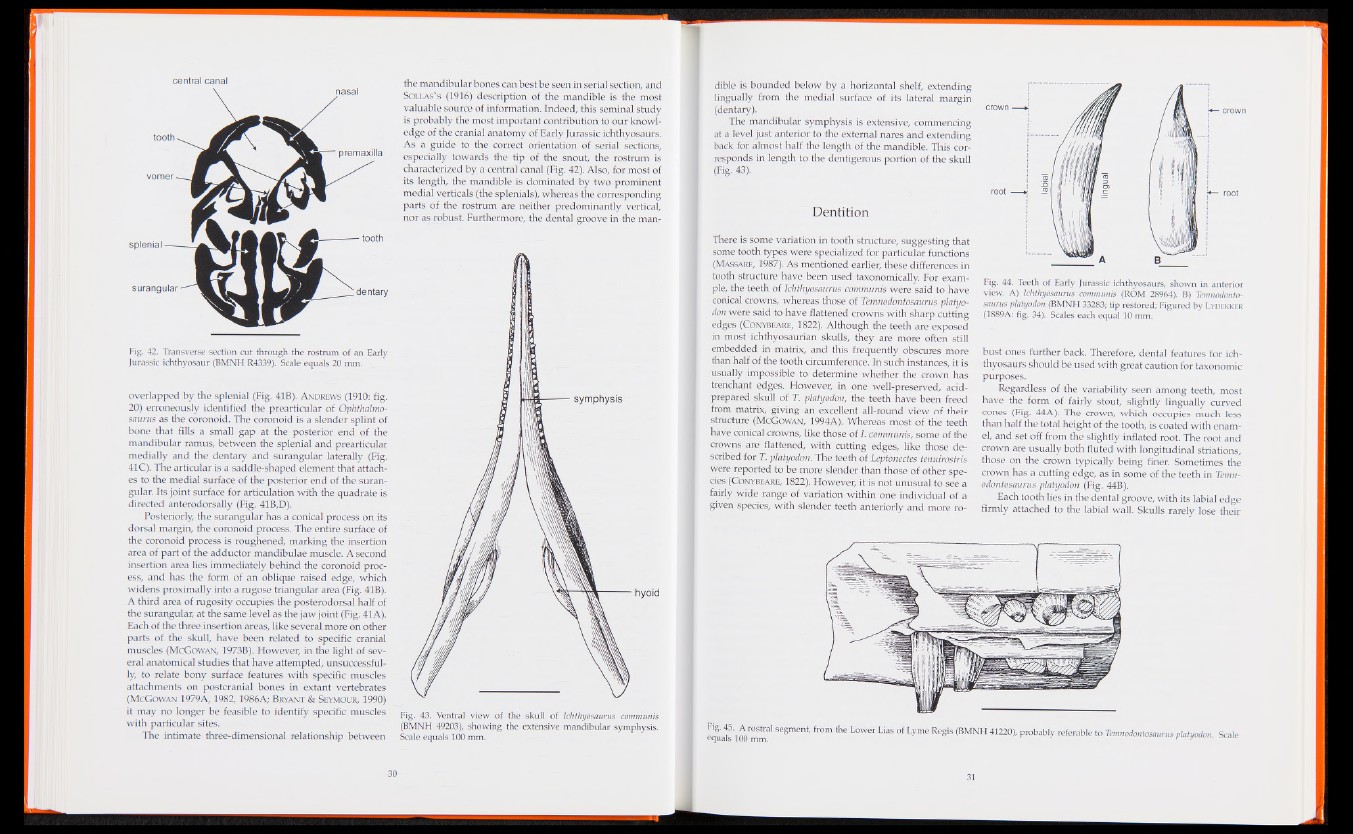
Fig. 42. Transverse section cut through the rostrum of an Early
Jurassic ichthyosaur (BMNH R4339). Scale equals 20 mm.
overlapped by the splenial (Fig. 41B). Andrews (1910: fig.
20) erroneously identified the prearticular of Ophthalmo-
saurus as the coronoid. The coronoid is a slender splint of
bone that fills a small gap at the posterior end of the
mandibular ramus, between the splenial and prearticular
medially and the dentary and surangular laterally (Fig.
41C). The articular is a saddle-shaped element that attaches
to the medial surface of the posterior end of the suran-
gular. Its joint surface for articulation with the quadrate is
directed anterodorsally (Fig. 41B,D).
Posteriorly, the surangular has a conical process on its
dorsal margin, the coronoid process. The entire surface of
the coronoid process is roughened, marking the insertion
area of part of the adductor mandibulae muscle. A second
insertion area lies immediately behind the coronoid process,
and has the form of an oblique raised edge, which
widens proximally into a rugose triangular area (Fig. 41B).
A third area of rugosity occupies the posterodorsal half of
the surangular, at the same level as the jaw joint (Fig. 41 A).
Each of the three insertion areas, like several more on other
parts of the skull, have been related to specific cranial
muscles (McGowan, 1973B). However, in the light of several
anatomical studies that have attempted, unsuccessfully,
to relate bony surface features with specific muscles
attachments on postcranial bones in extant vertebrates
(McGowan 1979A, 1982, 1986A; Bryant & Seymour, 1990)
it may no longer be feasible to identify specific muscles
with particular sites.
The intimate three-dimensional relationship between
the mandibular bones can best be seen in serial section, and
Sollas’s (1916) description of the mandible is the most
valuable source of information. Indeed, this seminal study
is probably the most important contribution to our knowledge
of the cranial anatomy of Early Jurassic ichthyosaurs.
As a guide to the correct orientation of serial sections,
especially towards the tip of the snout, the rostrum is
characterized by a central canal (Fig. 42). Also, for most of
its length, the mandible is dominated by two prominent
medial verticals (the splenials), whereas the corresponding
parts of the rostrum are neither predominantly vertical,
nor as robust. Furthermore, the dental groove in the man-
Fig. 43. Ventral view of the skull of Ichthyosaurus communis
(BMNH 49203), showing the extensive mandibular symphysis.
Scale equals 100 mm.
dible is bounded below by a horizontal shelf, extending
lingually from the medial surface of its lateral margin
(dentary).
The mandibular symphysis is extensive, commencing
at a level just anterior to the external nares and extending
back for almost half the length of the mandible. This corresponds
in length to the dentigerous portion of the skull
(Fig. 43).
Dentition
There is some variation in tooth structure, suggesting that
some tooth types were specialized for particular functions
(Massare, 1987). As mentioned earlier, these differences in
tooth structure have been used taxonomically. For example,
the teeth of Ichthyosaurus communis were said to have
conical crowns, whereas those of Temnodontosaurus platyo-
don were said to have flattened crowns with sharp cutting
edges (Conybeare, 1822). Although the teeth are exposed
in most ichthyosaurian skulls, they are more often still
embedded in matrix, and this frequently obscures more
than half of the tooth circumference. In such instances, it is
usually impossible to determine whether the crown has
trenchant edges. However, in one well-preserved, acid-
prepared skull of T. platyodon, the teeth have been freed
from matrix, giving an excellent all-round view of their
structure (McGowan, 1994A). Whereas most of the teeth
have conical crowns, like those of I. communis, some of the
crowns are flattened, with cutting edges, like those described
for T. platyodon. The teeth of Leptonectes tenuirostris
were reported to be more slender than those of other species
(Conybeare, 1822). However, it is not unusual to see a
fairly wide range of variation within one individual of a
given species, with slender teeth anteriorly and more roB
Fig. 44. Teeth of Early Jurassic ichthyosaurs, shown in anterior
view. A) Ichthyosaurus communis (ROM 28964). B) Temnodontosaurus
platyodon (BMNH 33283; tip restored; Figured by L ydekker
(1889A: fig. 34). Scales each equal 10 mm.
bust ones further back. Therefore, dental features for ichthyosaurs
should be used with great caution for taxonomic
purposes.
Regardless of the variability seen among teeth, most
have the form of fairly stout, slightly lingually curved
cones (Fig. 44A). The crown, which occupies much less
than half the total height of the tooth, is coated with enamel,
and set off from the slightly inflated root. The root and
crown are usually both fluted with longitudinal striations,
those on the crown typically being finer. Sometimes the
crown has a cutting edge, as in some of the teeth in Temnodontosaurus
platyodon (Fig. 44B).
Each tooth lies in the dental groove, with its labial edge
firmly attached to the labial wall. Skulls rarely lose their
e q t S ' Se8ment' &0m * e L°Wer LiaS ° f Lyme RegiS (BMNH 41220)' Probably re£erable to Temnodontosaurus platyodon. Scale