90 E X P L A N A T I O N OF THE TABLES.
E, Ligaments which connea the ventricle of the heart to the pericardium.
F G, The lower part of the cefophagus and upper part of the ftomach laid
open.
H, The under end of the ftomach.
I, The pylorus.
K, The beginning of a du£l from the lower end of the cefophagus to the airbag.
L M, A probe paiTed from the lower end of tl^at duct into O the air-bag.
N N, Two red fpheroidal bodies placed at the fides of the cnti-ance of the airdud
into tlie air-bag.
T A L E XVI.
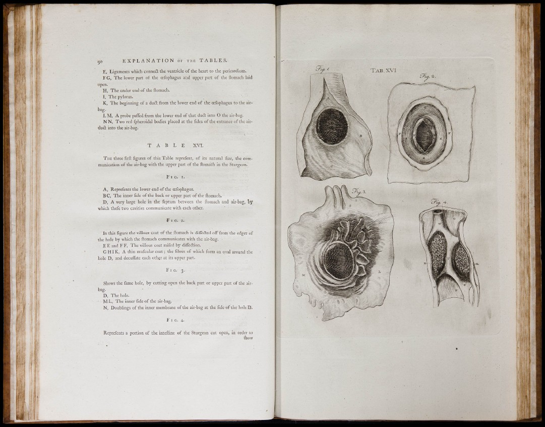
T H E three FIRFT figures of tliis Table reprefent, of its natural fize, the communication
of the air-bag witli the upper part of tlie ilomach in the Sturgeon.
F i g . r.
A, Reprefents the lower end of the cefophagus.
BC, The inner fide of the back or upper part of the ilomach.
D, A very large hole in the feptum between tlie ilomach and air-bag, by
which thefe two cavities communicate witli each odier.
In this figure the villous coat of the ilomach 5s diflecled off from the edges of
the hole by which the ilomach communicates with the air-bag.
E E and F F, The villous coat raifed by diiTedlion.
G H IK, A thin mufcular coat the fibres of which form an oval around the
hole D, and decuflate each other at its upper part.
F i g . 3.
Shows the fame hole, by cutting open the back part or upper part of tlie airbag.
D, The hole.
M L, The inner fide of the air-bag,
N, Doublings of the inner membrane of the air-bag at the fide of the hole D.
F I G. 4.
Reprefents a portion of the inteiline of the Sturgeon cut open, in order to
ihow
r s
' 1 1
I l :
I f
i - i l
i l i l :
]
1if1l1il ii