i ; il
ii
i j i i i - i i.
l i ' : " '
\ '-'If!
il
i ' l ' i
1% ^
I
l i :
I
Ï
' I t e - f f l . if'
I V
I f ]
E X P L A N A T I O N 0? ÌHE TABLES ,
clef. Three great branches into which the vena portar urn divides, and which
are difperfed l ike arteries in the liver for the fecretion o f the bile.
ghh, K faint iketch of the ladeals from the fmall inteftine.
i i i , Lafleals from the fundus o f thè ftomach joined b y lymphatics from the
f p l e e n and pancreas.
k, A plexus where the above-mentioned ladeals and lymphatics communicate.
//, Lymphatics from the fpl e en and ladleals from the lefler curvature of the
ftomach.
m, Lymphatics from the gall-bladder.
n «, Lymphatics from the liver.
0, La£leals from the- cefophagus.- •
pq, K large and intricate plexus of the trunks formed by all the abovem
e n t i o n e d lymphatics and iacteals, which runs upwards and forwards between
the cefophagus and fpine connef led to the celiac and mefenteric arteries.
li
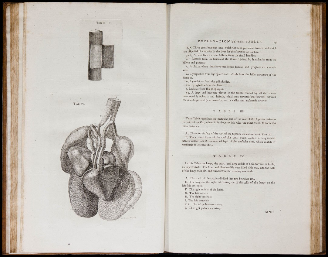
T A B L E II I * .
T H I S Table reprefents the mufcular coat of the root of the fuperior mefenter
i c vein of an O x , where it is about to joi n with the other veins, to form tlie
vena portarum.
A , The outer furface o f the root of the fuperior mefenteric vein of an ox.
B , The external layer of the mufcular coat, which confifts of longitudinal
fibres ; raifed from C , the internal layer of the mufcnlar coat, which confifts of
t r a n f v e r f e or circular fibres.
T A B L E IV .
IN this T abl e the lungs, the heart, and large veflels o f a fea-tortoifc or turtle,
arc reprefented. The heart and blood-veffels were filled with wax, and the cells
o f the lungs with air, and dried before the drawing was made.
A , The trunk o f the trachea divided into two branches B C.
D , The lungs on the right fide entire^ and E the cells of the lungs on the '
l e f t fide cut open.
F , The right auricle of the heart.
G , The left auricle.
H , The right ventricle.
I , The left ventricle.
K K, The left pulmonary artery.
T h e right pulmonary artery.
M N O ,