about an eighth the size of the larger,—the latter, however, being nearly equal, and their
nuclei contain several nucleoli.
, Some females retain a considerable number of ova till the middle of May, though
the body is by no means distended. Many have been discharged.
Fig. 27.
Besides the larger ova are many minute forms attached to the germinal tissue, so
that the spawning period is prolonged, or the minute ova retained or absorbed.
About the middle of February specimens with masses of ova under the scales are
common. The slightly pinkish eggs are attached to each other and the surface by a
transparent mucous secretion, so that they do not readily fall off. They .form a dense
layer under the scales, and in some are almost invisible. The process would appear to
be protective, giving them the shelter of the adult, and enabling them to escape the
attacks of predatory crustaceans or other forms. They have a diameter of about *56
to '78 mm.; the zona is delicate and translucent, yet resists some pressure. In the
perivitelline space are a few granular cells, such as those found in various eggs of fishes
(e. g. the gurnard). In structure the yolk is minutely granular, the figure showing the
Fig. 28.
Segmenting ovum of ïïarmothoë imbricata.
egg cleft into two spheres (Fig. 28). The eggs are so opaque that section alone reveals
the structure. In confinement the females carrying ova readily throw off their scales, a
feature probably due to the absence of nourishment and the condition of the water.
The males have their spermatozoa fully developed at the end of January and
beginning of February. These consist of simple tapering rods with a very attenuate
filament from the broader end (Plate XXVI a, fig. 1 ). They thus differ in shape from
those of Lepidonotus squa/matus (Plate XXVI a , fig. 2). When punctured the tissues of
a ripe male heal in a day or two, and the animal regains activity.
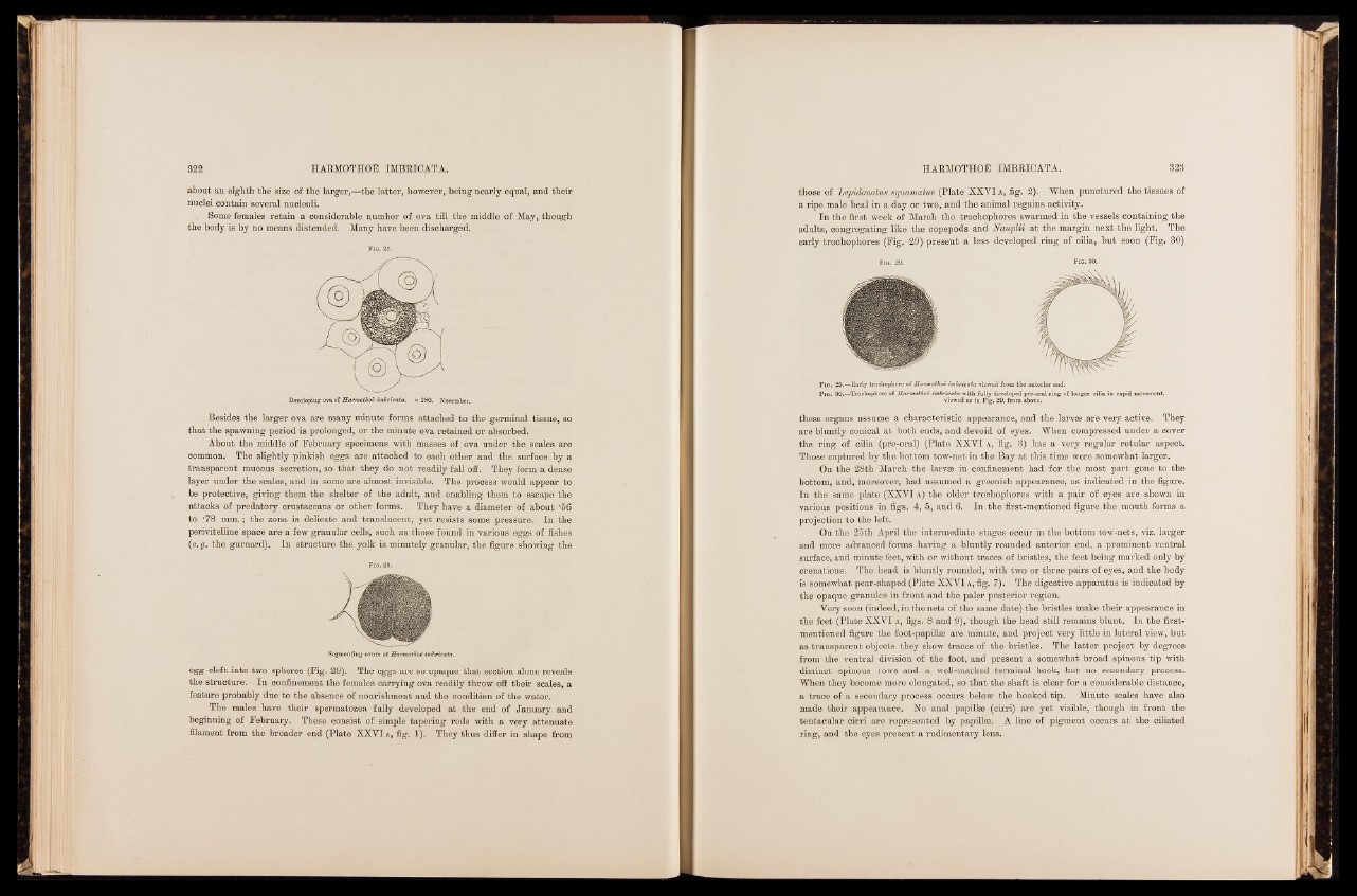
In the first week of March the trochophores swarmed in the vessels containing the
adults, congregating like the copepods and Nauplii at the margin next the light. The
early trochophores (Fig. 29) present a less developed ring of cilia, but soon (Fig. 30)
Fig. 29. Fig. 30.
these organs assume a characteristic appearance, and the larvae are very active. They
are bluntly conical at both ends, and devoid of eyes. When compressed under a cover
the ring of cilia (pre-oral) (Plate XXVI a , fig. 3) has a very regular rotular aspect.
Those captured by the bottom tow-net in the Bay at this time were somewhat larger.
On the 28th March the larvm in confinement had for the most part gone to the
bottom, and, moreover, had assumed a greenish appearance, as indicated in the figure.
In the same plate (XXVI a ) the older trochophores with a pair of eyes are shown in
various positions in figs. 4, 5, and 6. ' In the first-mentioned figure the mouth forms a
projection to the left.
On the 25th April the intermediate stages occur in the bottom tow-nets, viz. larger
and more advanced forms having a bluntly rounded anterior end, a prominent ventral
surface, and minute feet, with or without traces of bristles, the feet being marked only by
crenations. The head is bluntly rounded, with two or three pairs of eyes, and the body
is somewhat pear-shaped (Plate XXVI a , fig. 7). The digestive apparatus is indicated by
the opaque granules in front and the paler posterior region.
Very soon (indeed, in the nets of the same date) the bristles make their appearance in
the feet (Plate XXVI a , figs. 8 and 9), though the head still remains blunt. In the first-
mentioned figure the foot-papillae are minute, and project very little in lateral view, but
as transparent objects they show traces of the bristles. The latter project by degrees
from the ventral division of the foot, and present a somewhat broad spinous tip with
distinct spinous rows and a well-marked terminal hook, but no secondary process.
When they become more elongated, so that the shaft is clear for a considerable distance,
a trace of a secondary process occurs below the hooked tip. Minute scales have also
made their appearance. No anal papillae (cirri) are yet visible, though in front the
tentacular cirri are represented by papillae. A line of pigment occurs at the ciliated
ring, and the eyes present a rudimentary lens.