of a brownish hue. It is marked by the transverse ridges indicating the segments. The
arrangement of the folds at the mouth is seen in Plate I, fig. 1.
The papillae of the segmental organs commence at the ninth and continue to the
twenty-third foot. There thus appear to be fifteen pairs. Thé removal of the apertures
of these organs entirely from the ventral surface is a feature of the sub-family, and
distinguishes them at once from the Polynoidae.
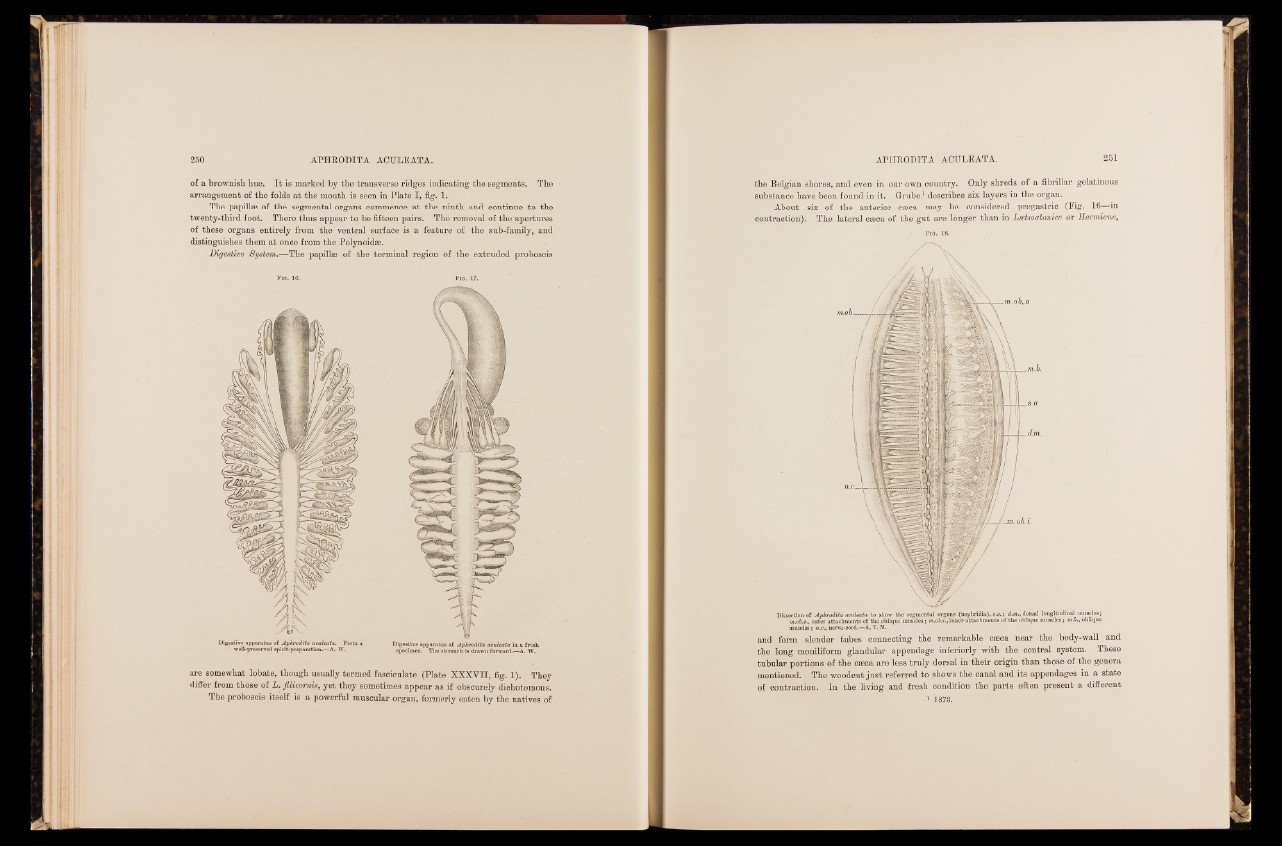
Digestive System.—The papillae of the terminal region of the extruded proboscis
Digestive apparatus of Aphrodita aculeata. From a
well-preserved spirit-preparation.—A. W.
Digestive apparatus of Aphrodita aculeata in a fresh
specimen. The stomach is drawn forward.—A. W.
are somewhat lobate, though usually termed fasciculate (Plate XXXVII, fig. 1). They
differ from those of L. filicomis, yet they sometimes appear as if obscurely dichotomous.
The proboscis itself is a powerful muscular organ, formerly eaten by the natives of
the Belgian shores, and even in our own country. Only shreds of a fibrillar gelatinous
substance have been found in it. Grube1 describes six layers in the organ.
About six of the anterior caeca may be considered pregastric (Pig. 16—in
contraction). The lateral caeca of the gut are longer than in Lsetmatonice or Hermione,
Fig. 18.
Dissection of Aphrodita aculeata to show the segmental organs (nephridia), s.o.; d.m., dorsal longitudinal muscles;
m.ob.o., outer attachments of the oblique muscles; m.ob.i., inner attachments o f the oblique muscles; m.b., oblique
muscles; ».c.,nerve-cord.—A.T.M.
and form slender tubes connecting the remarkable caeca near the body-wall and
the long moniliform glandular appendage inferiorly with the central system. These
tubular portions of the caeca are less truly dorsal in their origin than those of the genera
mentioned. The woodcut just referred to shows the canal and its appendages in a state
of contraction. In the living and fresh condition the parts often present a different
1873.