879.
1881.
1884.
1886.
1888.
1889.
1890.
1891.
1896.
1897.
1898.
Polynoë imbricata, Théel. Kong! sv. Yet. Akad. Handl., Bd. xvi, 3, p. 9.
,y cirrata, Horst. Niederland. Archiv Zool., 1881, Suppl. Bd. i, p. 5.
» » Pelsener. Bull. Soc. Roy. Malacol. Belg., xiv, p. lxxxis.
}, „ Kallenbach. Inaugur. Dissert. Eisenach, 1883. '
Harmothoë imbricata, Levinsen. Nord. Annulât., 194.
Polynoë imbricata, L. Wirén. Chætop. ‘Yoga* Exped., &c., p. 389.
,, cirrata, Carus. Eauna Médit., i, 201.
Harmothoë imbricata, Webster and Benedict. Ann. Mass., 701r
,} 33 Harvey Gibson. - Yerm. Liverp., 149.
» 33 De St. Joseph. Ann. Sc. Nat., 1888, p. 161, pl. vii, f. 21.
« 33 Trautzsch. Jenaische Zeitsch. f. Nat., xxiv, p. 66, and Arch. f.Naturges.,
55 Jahr, Bd. i, Hft. 2, p. 136, pl. vii, f. 1.
33 33 Malaquin. Ann. Boulon., 21.
Polynoë (.Harmothoë) imbricata, Homell. Polychæta, Liverpool Dist., p. 231, pl. xiii, f. 2. .
Harmothoë imbricata, Michaelsen. Polych. Fauna, p. 11.
Polynoë (Harmothoë) imbricata, Roule. Camp. d. ‘ Caud./ 443.
Harmothoë imbricata, H. P. Johnston. Pacific, Annel., Califor. Acad. Sc., 181, pl. vii, f. 37.
33 33 Michaelsen. Grönl., Ann., p. 121.
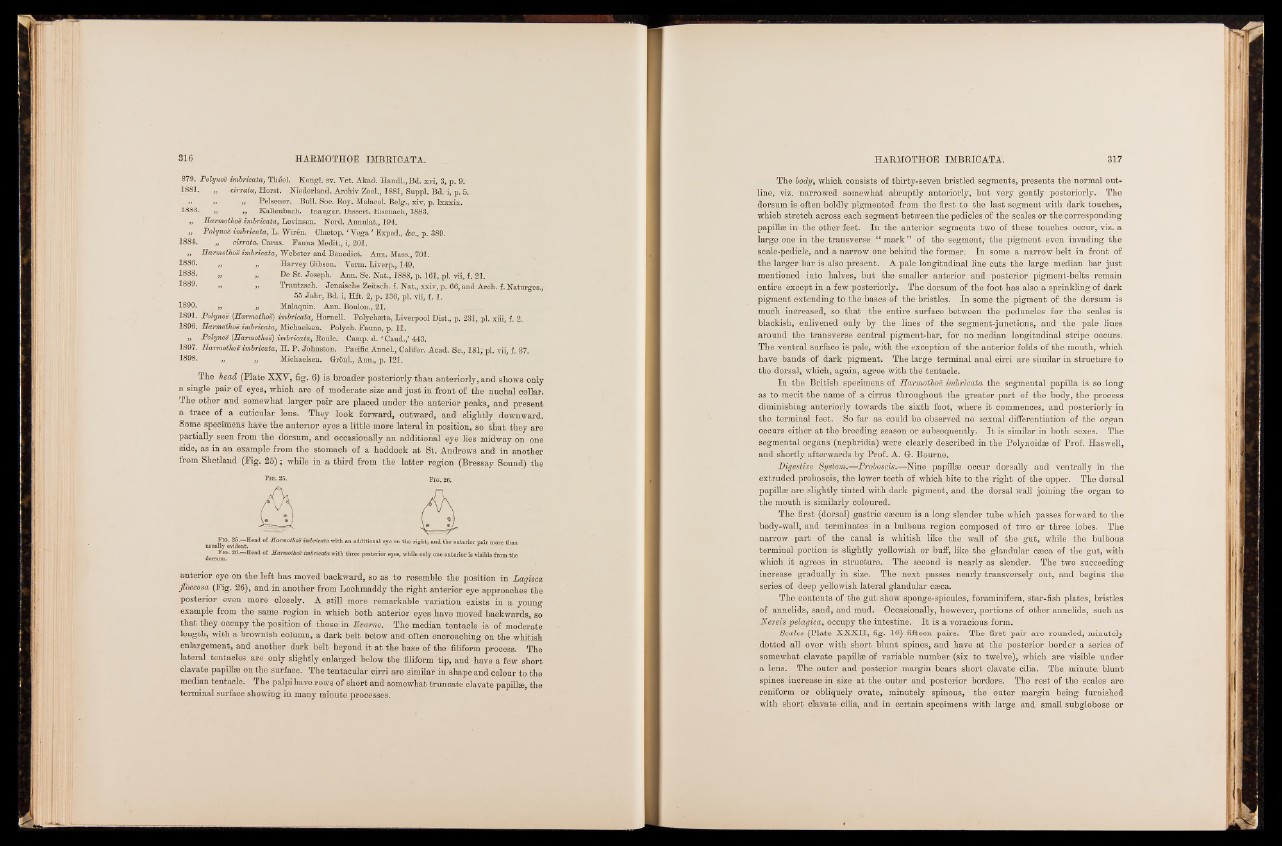
The head (Plate XXY, fig. 6) is broader posteriorly than anteriorly, and shows only
a single pair of eyes, which are of moderate size and just in front of the nuchal collar.
Ihe other and somewhat larger pair are placed under the anterior peaks, and present
a trace of a cuticular lens. They look forward, outward, and slightly downward.
Some specimens have the anterior eyes a little more lateral in position, so that they are
partially seen from the dorsum, and occasionally an additional eye lies midway on one
side, as in an example from the stomach of a haddock at St. Andrews and in another
from Shetland (Fig. 25); while in a third from the latter region (Bressay Sound) the
F ia . 25. Head of Harmothoe imbricata with an additional eye on the rig h t, and the anterior p air more than
usnally evident.
Fig. 26.—Head of Harmothoe imbricata with three posterior eyes, while only one anterior is visible from the
dorsum.
anterior eye on the left has moved backward, so as to resemble the position in Lagisca
fioccosa (Fig. 26), and in another from Lochmaddy the right anterior eye approaches the
posterior even more closely. A still more remarkable variation exists in a young
example from the same region in which both anterior eyes have moved backwards, so
that they occupy the position of those in Evame. The median tentacle is of moderate
length, with a brownish column, a dark belt below and often encroaching on the whitish
enlargement, and another dark belt beyond it at the base of the filiform process. The
lateral tentacles are only slightly enlarged below the filiform tip, and have a few short
clavate papillaa on the surface. The tentacular cirri are similar in shape and colour to the
median tentacle. The palpi have rows of short and somewhat truncate clavate papillse, the
terminal surface showing in many minute processes.
The body, which consists of thirty-seven bristled segments, presents the normal outline,
viz. narrowed somewhat' abruptly anteriorly, but very gently posteriorly. The
dorsum-is often boldly pigmented from the first to the last segment with dark touches,
which stretch across each segment between the pedicles of the scales or the corresponding
papillae in the other feet. In the anterior segments two of. these touches occur, viz. a
large one in the transverse “ mark” of the segment; the pigment even invading the
scale-pedicle, and a narrow one-behind the former. In some a narrow belt in front of
the larger bar is also present. A. pale longitudinal line cuts the large median bar just
mentioned into halves, but the smaller anterior and posterior pigment-belts remain
entire except in a few posteriorly. The dorsum of the foot has also a sprinkling of dark
pigment extending to the bases of the bristles. In some the pigment of the dorsum is
much increased, so. that the entire surface between the peduncles for the scales is
blackish, enlivened only by the lines of the segment-junctions, and the pale lines
around the transverse central pigment-bar, for no median longitudinal stripe occurs.
The ventral surface is pale, with the exception of the anterior folds of the mouth, which
have bands of dark pigment. The large terminal anal cirri are similar in structure to
the dorsal, which, again, agree with the tentacle.
In the British specimens of Harmothoë imbricata the segmental papilla is so long
as to merit the name of a cirrus throughout the greater part of the body, the process
diminishing anteriorly towards the sixth foot, where it commences, and posteriorly in
the terminal feet. /So far as'could be observed no sexual differentiation of the organ
occurs either at the breeding season or subsequently. It is similar in both sexes. The
segmental organs (nephridia) were clearly described in the Polynoidæ of Prof. Haswell,
and shortly afterwards by Prof. A. Gr. Bourne.
Digestive■ System.—Proboscis.-—Nine papillæ occur dorsally and ventrally in the
extruded proboscis, the lower teeth of which Bite to the right of the upper. The dorsal
-papillæ are slightly tinted with dark pigment, and the dorsal wall joining the organ to
the mouth is similarly coloured.
The first (dorsal) gastric cæcum is a long slender tube which passes forward to the
body-wall, and terminates in a bulbous region composed of two or three lobes. The
narrow part of the canal is whitish like the wall of the gut, while the bulbous
terminal portion is slightly yellowish or buff, like the glandular cæca of the gut, with
which it agrees in structure. The second is nearly as slender. The two succeeding
increase gradually in size. The next passes nearly transversely out, and begins the
series of deep yellowish lateral glandular cæca.
The contents of the gut show sponge-spicules, foraminifera, star-fish plates, bristles
of annelids, sand, and mud. Occasionally, however, portions of other annelids, such as
Nereis pelagica, occupy the intestine. I t is a voracious form.
Scales (Plate XXXII, fig. 10) fifteen pairs. The first pair are rounded, minutely
dotted all over with short blunt spines, and have at the posterior border a series of
somewhat clavate papillæ of variable number (six to twelve), which are visible under
a lens. The outer and posterior margin, bears short clavate cilia. The minute, blunt
spines increase in size at the outer and posterior borders. The rest of the scales- are
reniform or obliquely ovate, minutely spinous, the outer margin being furnished
with short clavate cilia, and in certain specimens with large and small subglobose or