riiiLc à x ià .
P l a t e XXIX.
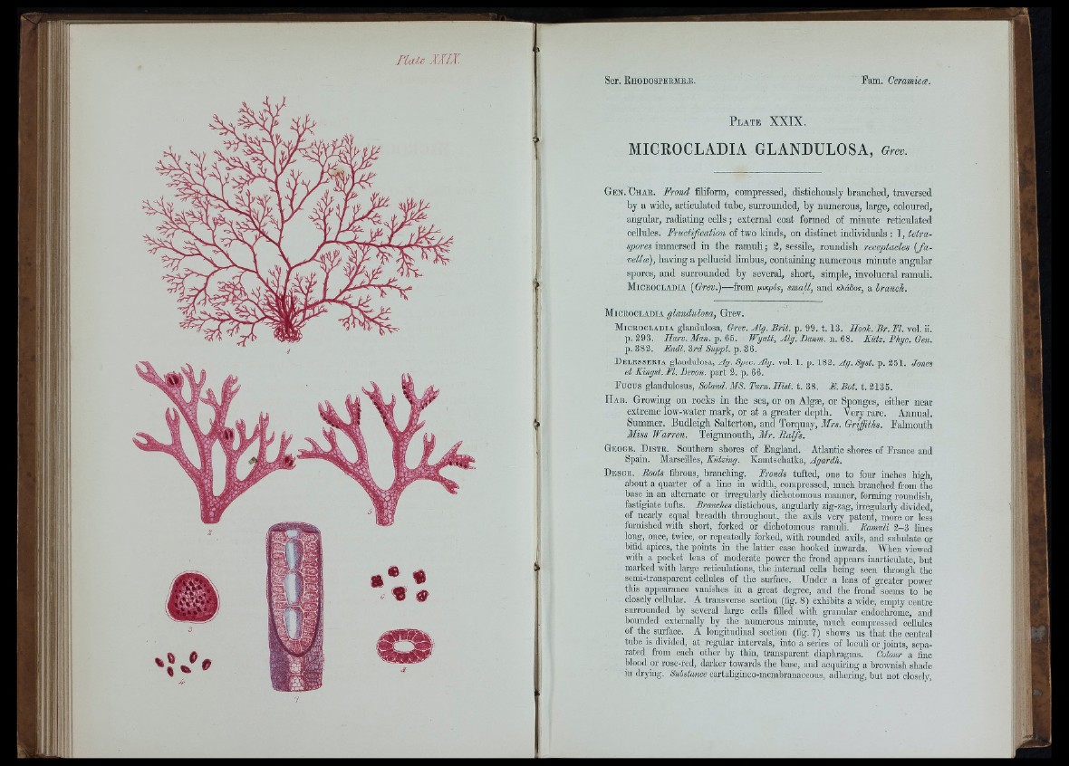
MICEOCLADIA GLANDULOSA, Greo.
Gen. Chab. Frond filiform, compressed, distioliously branched, traversed
by a wide, articulated tube, surrounded, by numerous, large, coloured,
angular, radiating cells; external coat formed of minute reticulated
ceUules. Fructification of two kinds, on distinct individuals : 1, tetra-
spores immersed in the ramuli; 3, sessile, roundish receptacles { fa vella),
having a pellucid limbus, containing numerous minute angular
spores, and surrounded by several, short, simple, involucral ramuli.
Micbooladia {Grev.)—-from giKpos, small, and icXdSos, a branch.
. 1 .13. Ilooh. Br. FI. vol. ii.
;. n. 68. Kiitz. Phyc. Gen.
iL p. 351. Jones
Micbooladia glandulosa, Grev.
Micbocladia glandulosa, Grev. Alg. Brit. p.
p. 393. Harv. Man. p. 65. Wyatt, Alg.
p. 383, Eudl.Zrd Suppl.-ÿ.33.
Delesseria glandulosa, Ag. Spec. Alg. vol. 1. p. 183. Ag.
et Kingst.M. Devon, part 3. p. 66.
Fu cu s glandulosus, Soland. MS. Turn. H ist. t. 38. F. Bot. t. 3135.
H a b . Growing on rocks in the sea, or on Algæ, or Sponges, either near
extreme low-water mark, or at a greater depth. Very rare. Annual.
Summer. Budleigh Salterton, and Torquay, Mrs. Griffiths. Falmouth
Miss Warren. Teignmouth, Mr. Ralfs.
Geogr. Distr. Southern shores of England. Atlantic shores of France and
Spain. Marseilles, Kiitzing. Kamtschatka, Agardh.
Descr. Roots fibrous, branching. Fronds tufted, one to four inches high,
about a quarter of a hne in width, compressed, much branched from the
base in an alternate or irregularly diohotomous manner, forming roundish,
fastigiate tufts. Branches distichous, angularly zig-zag, hregularly divided,
of nearly equal breadth throughout, the axils very patent, more or less
furnished with short, forked or dichotomous ramidi. Ramuli 3 -3 Hnes
long, once, twice, or repeatedly forked, with rounded axils, and subulate or
bifid apices, the points in the latter case hooked inwards. MTien viewed
with a pocket lens of moderate power the frond appears inarticidate, but
marked with large reticulations, the internal cells being seen tlu-ough the
semi-transparent cellules of the sm-face. Under a lens of greater power
this appearance vanishes in a great degree, and the frond seems to be
closely cellular. A transverse section (fig. 8) exlühits a wide, empty centre
Slu-rounded by several large cells filled with granular endoclu-ome, and
bounded externally by the numerous minute, much compressed cellules
of the sm-face. A longitudinal section (fig. 7) shows us that the central
tube is divided, at regular intervals, into a series of locuh or joints, separated
from each other by thin, transpai-ent diaplu-agms. Colour a fine
blood 01- rose-red, darker towards the base, aud acqumng a brownish slmde
in di-yiug. Substance cartaligiuco-iiicmhrauaccous, adhering, but not closely,