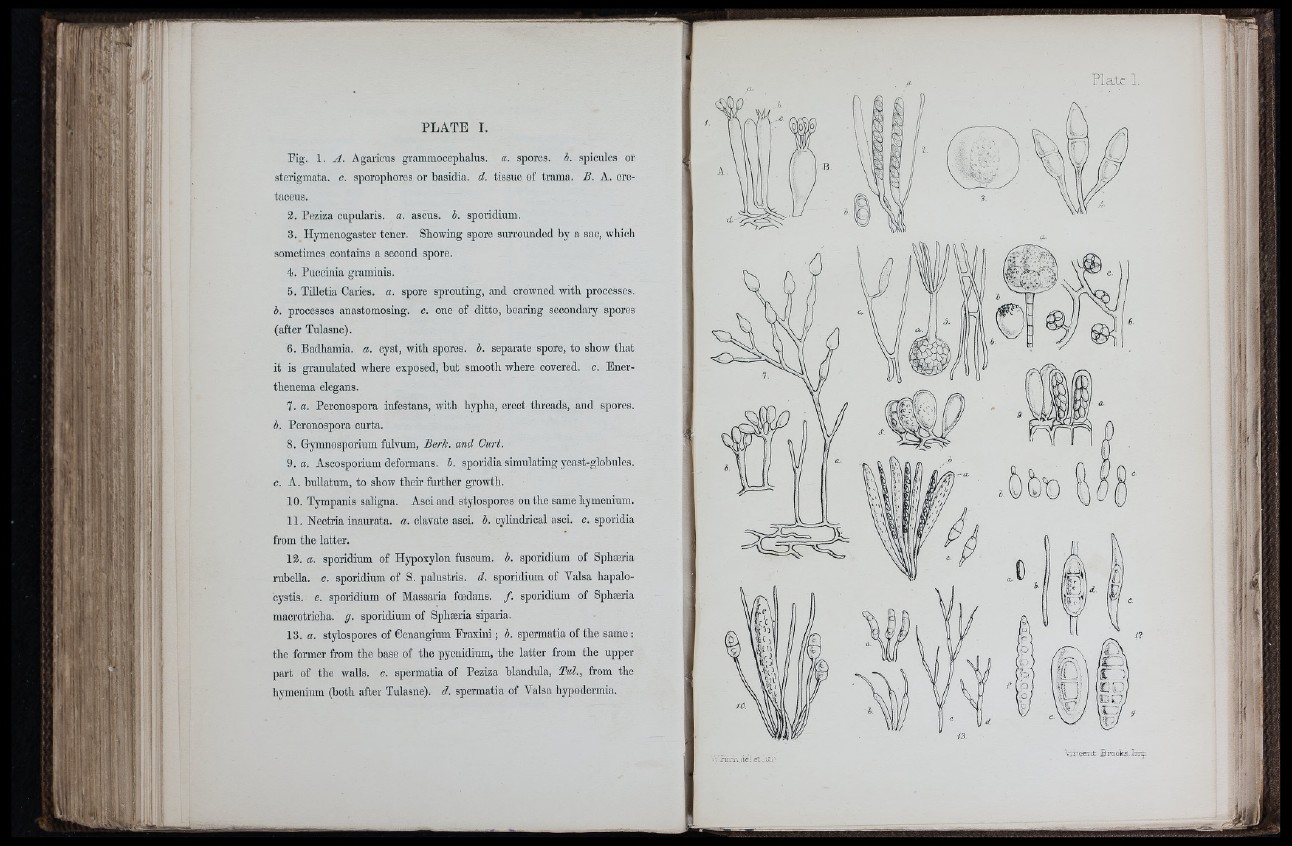
PLATE 1.
Fig. 1. A. Agaricus grammooeplialus. a. spores, i. spicules or
sterigmata. c. sporophores or basidia. d. tissue of trama. £. A. ore-
taceus.
2. Peziza cupularis. a. ascus. b, sporidium.
3. Hymenogaster tener. Showing spore surrounded by a sac, whicli
sometimes contains a second spore.
4. Puccinia graminis.
5. Tilletia Caries, a. spore sprouting, and crowned with processes.
b. processes anastomosing, c. one of ditto, bearing secondaiy spores
(after Tulasne).
6. Badhamia. a. cyst, with spores, b. separate spore, to show that
it is granulated where exposed, but smooth where covered, c. Ener-
thenema elegans.
7. a. Peronospora infestans, with hypha, erect threads, and spores.
b. Peronospora curta.
8. Gymnosporium fulvum. Berle, and Curt.
9.«. Ascosporium deformans, h. sporidia simulating yeast-globules.
c. A. bullatum, to show their further growth.
10. Tympanis saligna. Asci and stylospores on the same hymenium.
11. Nectria inaurata. a. clavate asci. h. cylindrical asci. c. sporidia
from the latter.
13. a. sporidium of Hypoxylon fuscum. b. sporidium of Sphæria
rubeUa. c. sporidium of S. palustris. d. sporidium of Valsa hapalocystis.
e. sporidium of Massaria foedans. f. sporidium of Sphæria
macrotricha. g. sporidium of Sphæria siparia.
13. a. stylospores of Cenangium Fraxini ; b. spermatia of the same ;
the former from the base of the pycnidium, the latter from the upper
part of the walls, c. spermatia of Peziza blandula, Tul., from the
hymenium (both after Tulasne). d. spermatia of Valsa hypodermia.
. riLV. aei.et-U
i i,' /rc ;:a
C l
CD muj
w j -
vntceiu Brooks
’ 1
! (I
1 n
ji . I
m