F R U C T I F I C A T I O N .
TAB. V I.
Anatomical views of the stigma and stigmatic surface of
B L E T I A T A N K E R V I L L I , E ; from a sketch by Mr. Bauer in
1802.
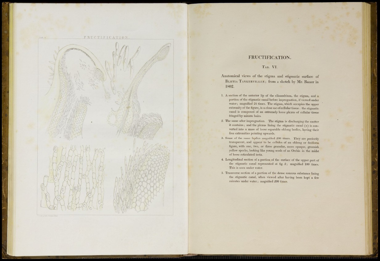
1. A section of the anterior lip of the clinandrium, the stigma, and a
portion of the stigmatic canal before impregnation, if viewed under
water; magnified 24 times. The stigma, which occupies the upper
extremity of the figure, is a close sac of cellular tissue : the stigmatic
canal is composed of an extremely loose plexus of cellular tissue
fringed by minute hairs.
2. The same after impregnation. The stigma is discharging the matter
it contains; and the plexus lining the stigmatic canal (a) is converted
into a mass of loose separable oblong bodies, having their
free extremities pointing upwards.
3. Some of the same bodies magnified 200 times. They are perfectly
transparent, and appear to be cellules of an oblong or fusiform
figure, with one, two, or three granular, more opaque, greenish
yellow specks, looking like young seeds of an Orchis in the midst
of loose reticulated testa.
4. Longitudinal section of a portion of the surface of the upper part of
the stigmatic canal represented at fig. 2; magnified 100 times.
This is seen under water.
5. Transverse section of a portion of the dense mucous substance lining
the stigmatic canal, when viewed after having been kept a few
minutes under water j magnified 200 times.