lied to, the preceding. The specific name is in allusion
to the slriking likeness in the form of the leaves, to
those o f Calophyllum Walkerii tab. 45 of the preceding
volume.
I am necessarily forced to pass over, unnoticed, many
species only known to me from description, often
not very perfect, but as my object is to sketch an arrangement,
it would tend to destroy its usefulness were i to
introduce species unknown to me as they might chance
to be placed in wrong sections, or might not even belong
to the genus. The following species I have ascertained
from the examination o f specimens must be excluded.
EXPLANATION OF
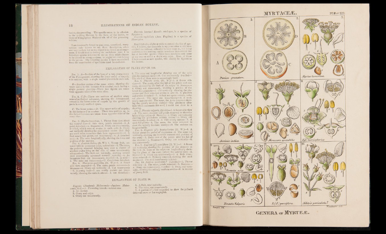
F ig. 1. A—Section of the base o f a very young ovary
o f the Pomegranate, showing the lower series of carpels,
4 in number, with a single central placenta in each cell.
B —Another section of the same ovary, showing the
upper series in this instance 6 in number, each having a
single parietal placenta—These two figures are taken
from opposite sides of the same slice.
F ig. 2. C.D—These are sections of another ovary
somewhat further advanced, showing, the derangement
caused in the lower series of carpels by the growth of
parts in a very confined space.
C. The lower series.—D . The upper series of carpels;
in this instance 7 in number. These two sections^ as in
the former instance are taken from opposite sides of the
same slice.
Fig. 3. Jfyrtus tomentosa. 1. Flower front view, about
the natural size—2. Side view, petals removed—&_A
petal detached—4. Stamens—5. Ovary cut transversely,
3-celied, with 2 rows of ovules in each, cell—6. Ovary
cut vertically showing the superposed ovules (but they
are much more numerous than here represented)—7. A
fruit nearly full grown, cut transversely—8. A detached
seed—9. The same longitudinally, showing the form and
position of the embryo.
Fig. 4. Jossinia indica, (R. W.) 1. Young fruit, covered
with its persistent calyx, natural size—2. The same,
the pericarp removed bringing into view a cluster of
aborted ovules lying on the side of some considerably
advanced—3. The fruit cut vertically, showing several
seed considerably advanced towards maturity—4. An
immature fruit cut transversely, 2-celled—5. A seed—
6. The same cut transversely—7. Cotyledons detached,
F ig. 5. Monoxora spectabilis, (R. W.)—1. A flower
side view, magnified—2. The same, petals removed—3.
Stamens—4. Ovary cut vertically—5. Cut transversely
—6. A young fruit—7. one nearly mature cut transversely,
showing the nuts in situ—fc. A nut detached—
EXPLANATION
Eugenia (Janibosa) Malaccensis-—Jambosa Malac-
censis, D.C.—1. Flowering branch—natural size.
2. An Anther.
3. Ovary and calyx.
4. Ovary cut transversely.
Eugenia laurina! Moon’s catalogue, is a species o f
Symptocos. " ■
Eugenia capitellata (Arn. Pugillus) is a species of
Memecylon.
Had I felt the slightest wish to extend the list of species,
I believe, the materials in my possession would have
enabled me without difficultyto have done so, but being
anxious on the contrary rather to reduce than extend the
list of doubtful species I have been careful to add none
of my own on which a doubt can exist. Most of those
I have named as new species, will shortly be figured in
the leones.
PLATE 97,* OR 122.
9. The same cut lengthwise showing one of the cells
with its enclosed seed—10. Cut transversely 3-celled—
The shell of these nuts is exceedingly hard.
Fig. 6. Pimenta acris, (R. W.)— 1. A flower side
view—2. The ovary,- after the fall of the petals and stamens,
crowned by the limb of the calyx—3. Stamens—
4. Ovary cut transversely, showing a portion of the
broad free placenta—5. Cut vertically showing the placentas
and their attached ovules in situ—6. A placenta
and ovules detached—7. A fruit not quite mature, natural
size—8. The same cut transversely—9. A seed,
nearly mature—10. The same, the testa removed showing
the spirally involute embryo—The glutinous albumen
which surrounds this seed, 1 could not show in a .
drawing. . . .
F ig. 7. Pimenta vulgaris, (Lind.)—1. A flower side view
__2. The same, stamens and petals removed to show the
lobes of .the calyx—3. Stamens—4. Ovary cut vertically
showing the pendulous solitary ovules—5. Ovary cut
transversely, 2-celled—6. A fruit nearly mature—7. A
seed full grown—8. The same, the testa removed to
show the spirally involute embryo—9. The embryo cut
longitudinally. ’
Fig. 8. Eugenia {/■) hemispheric (R. W.)—1. A
flower about the period of expansion—2. The same cut
vertically, showing the incurved stamens and position of
the ovary—3. Front view of the petals after the fall of
the stamens—4. Stamens—5. Ovary cut vertically—6.
cut transversely.
F ig. 9. Eugenia (J .) pauciflora (R. W.)—1. A flower
cut vertically, showing the position of the. stamens in
the bud—2. Tube of the calyx cut longitudinally showing
the place of the ovary (about the natural size)—3.
Stamens—4. Ovary cut transversely—5. A fruit somewhat
reduced-^6. Pericarp removed, showing the seed
in situ—7. The seed transversely 2-lobed.
Fig. 10. Nelitris paniculata? (Lindley)—1. Flower
side view—2. The same, petals removed—3. Stamens—
4. Ovary cut transversely, 8-celled—5. Ovary cut vertically,.
showing the solitary reniform ovules—6. A raceme
o f young fruit.
OF PLATE 98.
5. A fruit, near maturity.
6. The same, cut transversely.
7. A portion of a leaf magnified to show the pellucid
dots—all more or less magnified.
Ge n e r a of My r t e js .