G. Parsonie® ? Epigynum GriffUkanum (R. W.).
1. Corolla split open, stamens in situ. 2. Stamens.
3. Calyx, nectary, and pistil. 4. Ovary cut vertically,
adherent to the tube of the calyx. 5. Ovary cut
transversely.
H. Echiteae. Anadendron Candolianum[R.W.).
1. Expanded flower. 2. Detached corolla split open.
3. Calyx and stamens, after the removal of the corolla.
4. A detached stamen.
5. Two stamens and pistil. 6. Calyx cup-shaped,
nectary and pistil in situ. 7. Detached ovary. 8. The
same cut vertically.
9. Dissected seed, embryo in situ. 10. Detached
embryo, albumen sparing.
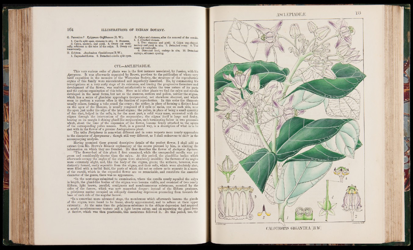
CVI.—ASCLEPIADE^E.
This very curious order of plants was in the first instance associated, by Jussieu, with his
Apocynece. I t was afterwards separated by Brown, previous to the publication of whose very
lucid exposition in the memoirs of the Wernerian Society, the structure of the reproductive
organs of this family were misunderstood and imperfectly described. He, by commencing his
investigations at a very early stage of its existence, and tracing the progressive formature and
development of the flower, was enabled satisfactorily to explain the true nature of its parts
and the curious organization of this tribe. Here as in other plants we find the calyx and corolla
developed in the usual forms, but not so the stamens, anthers and pollen, neither the stigma,
which has a series of glandular appendages (corpuscules), not elsewhere observed, and which
seem to perform a curious office in the function of reproduction. In this order the filaments
usually cohere, forming a tube round the ovary; the anther, in place of forming a distinct head
on the apex of the filament, is usually composed of 2 cells or sacks, one on each side, near
the apex just under the edge of the Inroad stigma; the pollen, in place of being a small quantity
of fine dust, lodged in the cells, is, for the most part, a solid waxy mass, connected with the
stigma through the intervention of the corpuscules; the stigma itself is large and fleshy,
bearing on its margin 5 shining gland-like corpuscules, each terminating below in two processes
which, about the time of the expansion of the flower, become firmly attached to the apices
of the corresponding pollen masses. Such, in a general way, is a description of what is to be
met with in the flower of a genuine Asclepiadeous plant.
The tribe Periphceae is somewhat different and in some respects more nearly approaches
to the character of Apocynaceae; though still very different, as I shall endeavour to show in the
accompanying analysis.
Having premised these general descriptive details of the perfect flower, I shall add an
extract from Mr. Brown’s Memoir explanatory of the course pursued by him, in eliciting the
information on which they are founded. He thus describes the flower of Asclepias Syriaca.
“The flower-bud of this plant I first examined, while the unexpanded corolla was yet
green and considerably shorter than the calyx. At this period, the gland-like bodies which
afterwards occupy the angles of the stigma were absolutely invisible; the furrows of its angles
were extremely slight, and, like the body of the stigma, green; the antheras, however, were
distinctly formed, easily separable from the stigma, and their cells, which were absolutely shut,
were filled with a turbid fluid, the parts of which did not so cohere as to separate in a mass;
of the cuculli, which in the expanded flower are so remarkable, and constitute the essential
character of the genus, there was no appearance.
“In the next stage submitted to examination, where the corolla nearly equalled the calyx
in length, the glandrlike bodies of the stigma were become visible, aud consisted of two nearly
filiform, light brown, parallel, contiguous and membranaceous substances, secreted by the
sides of the furrow, which was now somewhat deeper: instead of the filiform processes»
a gelatinous matter occupied an obliquely descending depression proceeding from towards the
base of each side of the angular furrow.
“In a somewhat more advanced stage, the membranes which afterwards become the glands
of the stigma, were found to be linear, closely approximated, and to adhere at their upper
extremity. At the same time the gelatinous substance in the oblique depression had acquired
a nearly membranaceous texture and a light brown colour, and on separating the gland from
it furrow, which was then practicable, this membrane followed it. At this period, too, the
ASCLEPIADEÆ
CAL0TR0PIS GIGANTEA[R.W)