ï
lo Mr D r um m o n d , nurseryman at I ’orfar, Avhom I have
already had occasion to notice as a most deserving and indefatigable
botanist, the British Flora is indebted for the discovery
of native specimens of this highly interesting species. It is
scarcely to be doubted, that his are the first which have been
detected in this country, though H e d w ig , in his Species
Muscorum, gives England as a general station without a question.
The same author, however, in his prior work, the Mus-
corumfrondosorum descriptio et adunbratio, says he received
a specimen from D ick so n , hut was ignorant whence he procured
it. No additional particulars are before the public.
I t is a widely distributed moss, having been found in most
parts of Europe, at the Cape of Good Hope, and in North
America. I have also received specimens from my friend D i-
R ic h a r d so n , collected in his journey to the Polar Sea, and
from the late Dr W r ig h t , gathered in Jamaica. The latter
specimens are remarkable for the cauline leaves possessing
more constantly a short nerve, than the European ones. In
Dr R ic h a r d so n ’s plants, I also observed a nerve reaching
half-way up some of the perichætial leaves. The nerve is indeed
a very fallacious character in several of the distichousleaved
Neckeroe, being present in some, and nearly absent in
other leaves on the same individual.
The acute M ohr seems to be^ the only museologist who
has described the leaves as serrulate at the apex. B r id e l , on
the other hand, observes : “ Semper autem integerrima contra
M o h r ium video.” The leaves of my specimens, however,
which were delineated before I was aware of the above discrepancy,
certainly are distinctly serrulate at the apex, as well as
in those from Jamaica, and those received from Dr R ic h a r d so
n . The inner peristome of N . pennata, is extremely fragile
and evanescent, and apt to escape very minute observation.
I J
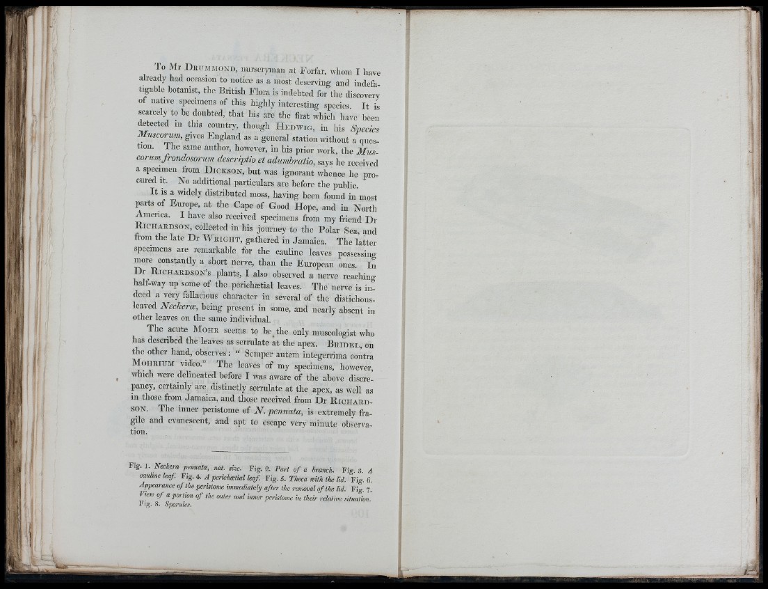
Fig. 1, Neckera pennata, nat. size. Fig. 2. Part o f a branch. Fig. 3. A
cauhne leaf. Fig. 4. A perieha:tial leaf. Fig. 5. Theca with the lid. Fig. 6.
Appearance o f the peristome immediately after the removal o f the lid. Fig. 7.
View o f a portion o f the outer and inner peristome in their relative situation.
Fig. 8. S])orules.
A