From the second cirrus a ridge passes as in other forms ventrally on each side. In
this species the anterior margin is 4—5-dentate, whilst in the centre is a deep hiatus.
In small examples the processes are slender tapering papillae. The branchiae on the next
two segments are typical. Large cement-glands, homologous with those of Lagis Koreni,
occur in the fifth and sixth segments. The duct opens behind the second branchia and
the first bristle-tuft. No muscle is present (Hessle).
The second region corresponds with that in other forms, viz., has more slender
bristles in smaller tufts than the succeeding. Their structure, however, corresponds
with the type common to all, including the posterior series (Plate CXXV, figs. 7—-7 c).
The stout simple bristles are tapered distally and have traces of wings (fig. 7 a). The
others have a spear-head enlargement at the end of the shaft and a tapering tip (fig. 7 b),
but the enlargement is proportionally broader and the tapered tip shorter than in allied
forms. In the posterior region the fourteen pairs of bristle-bundles exhibit a gradation
from the anterior to the posterior extremity. Moreover the region is only a little tapered
posteriorly, the termination being comparatively broad. In consequence the caudal
appendix projects little ventrally from the truncated end of the body, the last foot being
modified into a rounded flattened lobé projecting beyond the truncated surface and with
a subulate cirrus at its extremity, whilst the somewhat long row of caudal hooks is
intimately associated with its dorsal edge. No other hook or bristle is connected with it.
The dense rows of hooks are situated on the edge of the prominent lamellse.
Each has a short base or shaft (Plate CXXV, fig. 7 e) with a well-marked rounded crown,
a smaller and a larger fang beneath, the curve below the latter sloping to a modified
tooth with a 4—6-spinous edge, then a gulf below, and a rounded prow, the basal line
being slightly sinuous.1
The caudal appendix (scapha) presents dorsally an almost evenly truncated edge in
a line with the general surface (Plate CXXV, figs. 7 ƒ and 7 g), the margin, however, being
minutely crenulate and projecting a little beyond the dorsal surface of the appendix.
Then follows the line of caudal hooks, which abut at their ventral edge on the rounded
and flattened lamella with the cirrus. A notch separates the ventral edge of the lamella
from a series of four fimbriæ between it and the vent, the lower edge of which is crenate
with a subulate median cirrus. Nilsson2 has recently shown the structure.of the eyes of
this organ. The caudal hooks (Plate CXXV, fig. 7 d) are slightly narrowed at the base
of the striated shaft, then dilate, continue for some distance with nearly parallel sides,
diminish toward the neck, and end in a slight curvature at the point, which is somewhat
blunt, probably from friction.
The tube is slightly curved, and in Malmgren’s examples was composed of minute
shells, viz., Bissoa striata, and Bulla, truncata. Tubes from the coast of Kerry are composed
of comparatively large fragments of shells and stones with a minute Bissoa. Those from
422 fathoms off Ireland in the “ Porcupine ” Expedition of 1869 were formed of proportionally
large translucent grains of quartz with here and there a yellow and black grain of other
material. One fragmènt is composed of Foraminifera with a few grains of sand, but its
1 Malmgren figures the spinous edge as a simple process, whilst Hessle gives it six to seven teeth.
2 Beiträge Nervensyst. Polych. f Zool. Bidrag Uppsala/ Bd. i, p. 137, 1912.
identity is uncertain. A tube from 567 fathoms in the Atlantic in the “ Porcupine *
Expedition of 1870 presents a uniform series of dull yellow grains throughout. The
rounded and comparatively large yellow stones forming a tube from a depth of
52-^ fathoms (log 33) off the south west of Ireland are noteworthy.1
Family XXVIII.—Amphaiietidj5, Malmgren, 1867.
Cephalic lobe (upper lip of some) covering the mouth, the median part separated
by oblique grooves; tentacles long, smooth, pinnate or ciliated, arising from the
mouth, and can be engulfed. Buccal segment surrounding the mouth and forming
the lower' lip, occasionally biannulate. Tentacular membrane is divided by two longitudinal
grooves into three. A groove between tentacles and mouth. Body somewhat
broad in front, tapered posteriorly and with a variable number of segments, generally
twenty to forty, rarely about seventy, and of two regions, the anterior (or thoracic)
having fascicles of capillary bristles and pinnules for hooks, the posterior (or
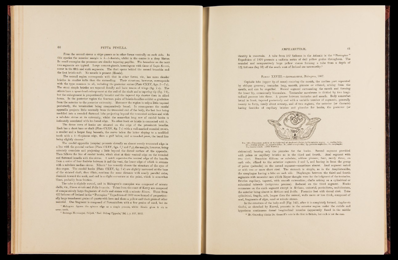
Fig. 143—Transverse section of the body-wall of Ampharete Grubei, in the anterior Gl.v., ventral glands; La., anterior lobes; Na., anterior nephridia; Np., posterior nreepghiorind,i a;e xO., ec.,a ordesiaocp hbaogduys;;
Vv., ventral vessel. (After Fauvel.)
abdominal) bearing only the pinnules for the books,. Seoond segment provided
with paleae or capillary bristles as in the third and fourth. Anal segment with
two cirri. Branchiae filiform or subulate, seldom pinnate, four, rarely three, on
each side, affixed to the anterior segments 2 and 3, and having in front the group
bf paleae (palmulse) on the Second segment—sometimes absent. Anal segment naked
or with two or more short cirri. The stomach is simple, as in the Amphictenidae,
the oesophagus having a lobe on each side. Diaphragm between the third and fourth
segments with muscular sacs which Meyer thought were for the lodgment of the tentacles.
Bristles capillary, tapered, with smooth extremities; shafts arising on a cylindrical or
subcorneal tubercle (setigerous . process). Reduced on the third segment. Hooks
commence on the sixth segment except in Melinna, uniserial, peetiniform, multidentate,
the anterior being absent in Melinna and Isolda. Posterior feet with dorsal cirri. Tube
cylindrical, fragile, soft, longer than the animal, walls more or less thick, composed of
mud, fragments of algae, sand or minute stones.
In the structure of the body-wall (Fig. 143), after it is completely formed, Ampharete
Qntbei, as sketched by Fauvel, presents, in the anterior region under the cuticle and
hypoderm continuous dorsal longitudinal muscles (apparently fused in the middle
1 Mr. Crawshay thinks Dr. Geramill’s note is the first in Britain, but such is’not the case.