of the blades touch the bases of the outer pale* and form a very regular second row, the
two sides making an ovoid ,area.
The inner or third row of the pale golden pale* forms an oblique palisade, which
leaves only a narrow ellipse between them, and in lateral view, in Neapolitan examples
especially, the palisade shows a high dorsal margin and diminishes gradually to the
ventral edge.v The typical palea (Plate CXXIII, fig. 3 b) has a long, flattened, tapering
terminal blade, from which the shaft passes off at an oblique angle and tapers to a point,
the heel or shbulder being at the front edge, the outline of which is very slightly concave,
and with serrations on the margin. The dorsal outline presents a slight convexity in the
region corresponding to the arch of the foot. The transverse or slightly oblique stri*
pass from the inner outline to the free edge where the notches are. The rest of the tip
is longitudinally striated.
By rendering the tissues transparent Arnold Watson1 has shown that in each
opercular lobe there are two setigerous sacs running longitudinally, the outer supplying
the outer pale*, the inner furnishing the median and inner pale* which lie alternately
in the sac. The developing pale* originate as minute, angular particles (cells?), and
the more advanced travel through the tissue in a somewhat spiral fashion to reach their
positions at the dorsal end of each opercular crescent. Thus new pale* take the places
of those which are injured or shed. The same observer counted the pale* in a large
example about 2 inches in length; . thus in the outer row on the left thirty-five
occurred, besides one just coming into view, thirty-six on the right and one just
visible; in the median row twenty-four on the left (three unpaired) and twenty-five
on the right, whilst the inner series consisted of twenty-one on the left and twenty on
the right.
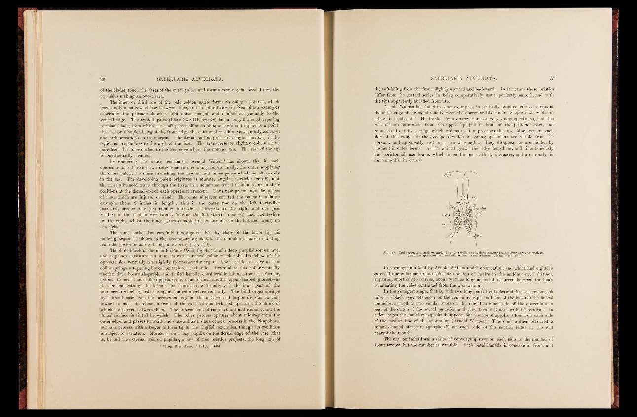
The same author has carefully investigated the physiology of the lower lip, his
building organ, as shown in the accompanying sketch, the strands of muscle radiating
from the posterior border being noteworthy (Fig. 138).
The dorsal arch of the mouth (Plate CXII, fig. 4 a) is of a deep purplish-browrn hue,
and it passes backward till it meets with a buccal collar which joins its fellow of the
opposite side ventrally in a slightly spout-shaped margin. From the dorsal edge of this
collar springs a tapering buccal tentacle on each side. External to this collar ventrally
another dark brownish-purple and frilled lamella, considerably thinner than the former,
extends to meet that of the opposite side, so as to form another spout-shaped process—as
it were ensheathing the former, and connected externally with the inner base of the
bifid organ which guards the spout-shaped aperture ventrally. The bifid organ springs
by a broad base from the peristomial region, the massive and larger division curving
inward to meet its fellow in front of the external spout-shaped aperture, the chink of
which is observed between them. The anterior end of each is blunt and rounded, and the
dorsal surface is tinted brownish. The other process springs about midway from the
outer edge, and passes forward and outward as a short conical process in the Neapolitan,
but as a process with a longer filiform tip in the English examples, though its condition
is subject to variation. Moreover, on a long papilla on the dorsal edge of the base (that
is, behind the external pointed papilla), a row of fine bristles projects, the long axis of
1 ‘Rep< Brit. Assoc.,’ 1910, p. <534.
the tuft being from the front slightly upward and backward. In structure these bristles
differ from the ventral series in being comparatively stout, perfectly smooth, and with
the tips apparently abraded from use.
Arnold Watson has found in some examples “ a centrally situated ciliated cirrus at
the outer edge of the membrane between the opercular lobes, as in 8. spimtlosa, whilst in
others it is absent.” He thinks, from observations on very young specimens, that this
cirrus is an outgrowth from the upper lip, just in front of the posterior part, and
connected to it by a ridge which widens as it approaches the lip. Moreover, on each
side of this ridge are the eye-spots, which in young specimens are visible from the
dorsum, and apparently rest on a pair of ganglia. They disappear or are hidden by
pigment in older forms. As the animal grows the ridge lengthens, and simultaneously
the peristomial membrane, which is continuous with it, increases, and apparently in
some engulfs the cirrus.
F ig. 138.—Oral region of a small example (} in.) of Sabellaria alveolata showing the building glandular apertures; in., muscular bands. From a sketch by Arnold Watsono. rgan bo., with its
In a young form kept by Arnold Watson under observation, and which had eighteen
external opercular pale* on each side and ten or twelve in the middle row, a distinct,
unpaired, short ciliated cirrus, about twice as long as broad, occurred between the lobes
terminating the ridge continued from the prostomium.
In the youngest stage, that is, with two long buccal tentacles and three others on each
side, two black eye-spots occur on the ventral side just in front of the bases of the buccal
tentacles, as well as two similar spots on the dorsal or inner side of the operculum in
rear of the origin of the buccal tentacles, and they form a square with the ventral. In
older stages the dorsal eye-specks disappear, but a series of specks is found on each side
of the median line of the operculum (Arnold Watson). The same author observed a
comma-shaped structure (ganglion?) on each side of the central ridge at the end
nearest the mouth.
The oral tentacles form a series of converging rows on each side to the number of
about twelve, but the number is variable. Each basal lamella is concave in front, and