as in other Mollusca, the whole body-cavity is filled with hsemolymph or blood-fluid, so
that the blood-spaces and blood-vessels may be regarded as merely divisions or diverticula
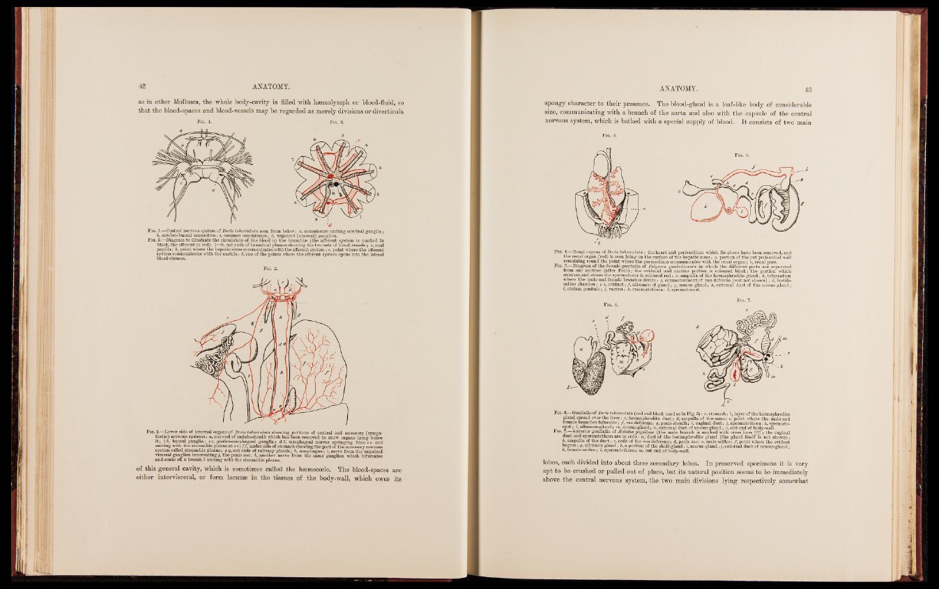
F ig. 1. Fig. 3.
F ig. 1.—Central nervous system of Doris tuberculata seen from below: a, commissure uniting cerebral ganglia;
b, cerebro-buccal connective; c, common commissure; d, unpaired (visceral) ganglion.
Fig. 3.—Diagram to illustrate the circulation of the blood in the branchiae (the afferent system is marked in
black, the efferent in red): 1—9, cut ends of branchial plumes showing the two sets of blood-vessels; a, anal
papilla; b, point where the hepatic sinus communicates with the afferent system; c, point where the efferent
system communicates with the auricle; d, one of the points where the efferent system opens into the lateral
blood-sinuses.
F ig. 2.
F ig. 2.—Dower side of internal organs of Doris tuberculata showing portions of central and accessory (sympathetic)
nervous systems: a, cut end of radula-sheath which has been removed to show organs lying below
i t ; b b, buccal ganglia; c c, gastro-oesophageal ganglia ; d d, oesophageal nerves springing from c c and
uniting with the stomachic plexus a t e e; ƒƒ, under side of stomach showing the part of the accessory nervous
system called stomachic plexus; g g, cut ends of salivary glands; h, oesophagus; i, nerve from the unpaired
visceral ganglion innervating j, the penis sac; k, another nerve from the same ganglion which bifurcates
and sends off a branch l uniting with the stomachic plexus.
of this general cavity, which is sometimes called the hmmocoele. The blood-spaces are
either intervisceral, or form lacunae in the tissues of the body-wall, which owes its
spongy Character to their presence. The blood-gland is a. leaf-like body of considerable
size, communicating with a branch of the aorta and also with the capsule of the central
nervous system, which is bathed with a special supply of blood. I t consists of two main
F ig. 4.
F ig. 4.—Renal organs of Doris tuberculata; the heart and pericardium which lie above have been removed, and
the renal organ (red) is seen lying on the surface of the hepatic mass; a, portion of the cut pericardial wall
remaining round the point where the pericardium communicates with the renal organ; b, renal pore.
F ig. 5.—Diagram of the female genitalia of Polycera quadrilineata in which the different parts are separated
from one another (after Pohl); the oviducal and uterine portion is coloured black; the portion which
receives and stores the spermatozoa is coloured red: a, ampulla of the hermaphrodite gland; b, bifurcation
where the male and female branches divide: c, commencement of vas deferens (rest not shown) ; d, fertilization
chamber; e e, oviduct; ƒ, albumen of gland; g, mucus gland; h, external duct of the mucus gland;
», atrium genitale; j, vagina; k, spermatotheca; l, spermatocyst.
Fig. 6. Genitalia of Doris tuberculata (red and black used as in Fig. 5) : a, stomach ; b, layer of the hermaphrodite
gland spread over the liver; c, hermaphrodite duct ; d, ampulla of the same; e, point where the male and
female branches bifurcate; ƒ, vas deferens; g, penis sheath; i, vaginal duct; j, spermatotheca; k, spermato-
cyst ; l, albumen-gland ; m, mucus-gland ; n, external duct of mucus-gland ; o, cut end of body-wall.
F ig. 7.—Anterior genitalia of Æolidia papillosa (the male branch is marked with cross lines = = ; the vaginal
duct and spermatotheca are in red) : a, duct of the hermaphrodite gland (the gland itself is not shown) ;
b, ampulla of the duct ; c, coils of the vas deferens ; d, penis sac; e, male orifice ; ƒ, point where the oviduct
begins ; g, albumen gland ; h, a portion of the shell-gland ; i, mucus-gland ; j, external duct of mucus-gland ;
■ k, female orifice ; l, spermatotheca ; m, cut end of body-wall.
lobes, eacb divided into about three secondary lobes. In preserved specimens it is very
apt to be crushed or pulled out of place, but its natural position seems to be immediately
above the central nervous system, the two main divisions lying respectively somewhat