PLATE I.
F igs.
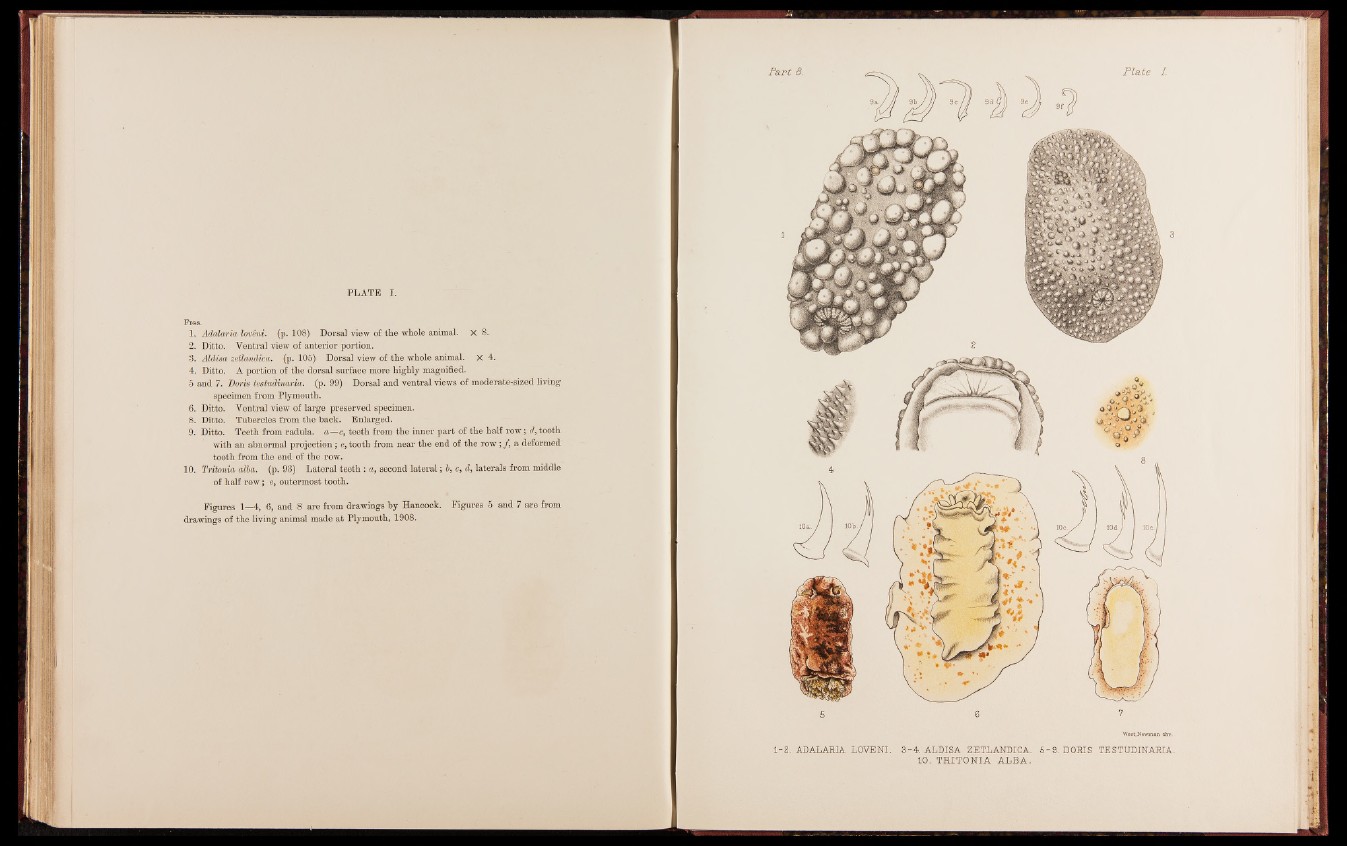
1. Adalaria loveni. (p. 108) Dorsal view of the whole animal. X 8.
2. Ditto. Ventral view of anterior portion.
3. Aldisa zetlandica. (p. 105) Dorsal view of the whole animal. X 4.
4. Ditto. A portion of the dorsal surface more highly magnified.
5 and 7. Doris testudinaria. (p. 99) Dorsal and ventral views of moderate-sized living
specimen from Plymouth.
6. Ditto. Ventral view of large preserved specimen.
8. Ditto. Tubercles from the back. Enlarged.
9. Ditto. Teeth from radula. a—c, teeth from the inner part of the half row; d, tooth
with an abnormal projection; e, tooth from near the end of the row; ƒ, a deformed
tooth from the end of the row.
10. Tritonia alba. (p. 93) Lateral teeth : a, second lateral; 6, c, d, laterals from middle
of half row; e, outermost tooth.
Figures 1—4, 6, and 8 are from drawings by Hancock. Figures 5 and 7 are from
drawings of the living animal made at Plymouth, 1908.
5 6
1 -2 . ADALARIA LOVENI. 3 - 4 . ALDISA ZETLANDICA. 5
10. T R IT O N IA A L B A .
-9 . DORIS TESTUDINARIA.