separated from each other, from the hook-rows. Internally is a thick coating of sarco-
lemma, but only a few distinct muscular fibres. The ventral longitudinal muscles form
ovoid masses often pointed internally in transverse section, and have internally the sarco-
lemma and muscular fibres continued from the dorsal. A wide gap separates them in
the middle line. The nerve-cords are placed in the substance of each near the upper
and inner border. This position removes them from contact with both the circular coat
and the hypoderm, and also from the influence of the oblique muscles, which are well
developed, and in an example of 8. alveolata observed by Mr. Arnold Watson a slip
passed from one oblique muscle to the adjoining one.
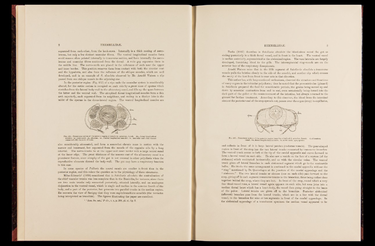
In the posterior region (Fig. 137) of a ripe male the muscular system is considerably
altered, for the entire coelom is occupied on ^ach side by a great mass of sperms which
stretches from the lateral body-wall to the alimentary canal, and fills up the space between
the latter and the ventral wall. The atrophied dorsal longitudinal muscles form a thin
arch superiorly, each separated from its neighbour, and ending in a thicker lobe in the
midst of the sperms in the dorso-lateral region. The ventral longitudinal muscles are
F ig. 136.—Transverse section of the anterior region of Sdbellaria spinulosa,muscles; nc., nerve-cord; pb., pharynx; vm., ventral longitudinal muscles ;L heui.c, km. usdcmul.a, rd oarnsdal oltohnegri ttuisdsiuneasl connected with the rows of hooks.
also considerably attenuated, and form a somewhat clavate mass in section with the
narrow end innermost, but separated from the muscle of the opposite side by a long
interval. The nerve-trunks lie at the upper and inner border with a large neural canal
at the inner edge. The great thickness of the mucous coat of the alimentary canal is a
prominent feature, since atrophy of the gut is not unusual in other polychaets when the
reproductive elements distend the body-wall. The gut may have a respiratory function-
in this case.
In some species of Pallasia the neural canals are smaller in front than in the
posterior region, and this raises the question as to the physiology of these structures.
Milne-Edwards1 (1838) considered that in Sdbellaria alveolata the centralisation of
the chief vascular trunks was less complete than in the Eunicidae, for instance, since there
are two main trunks only connected posteriorly, situated laterally, and an analogous
disjunction in the ventral vessel, which is single and median in the anterior fourth of the
body, and a part of the posterior, but presents two parallel trunks in the median region.
He corrects the view of Savigny that they were capitobranchiate annelids (the tentacles
being interpreted as branchiae). The figures illustrating the paper are excellent.
1 ‘ Ann. Sc. nat./ 2e ser., t. x, p. 208, pi. xi, fig. 3.
Fuchs (1907) describes in Sdbellaria alveolata the blood-sinus round the gut as
ending posteriorly in a thick dorsal vessel, and in front in the heart. The ventral vessel
is median anteriorly, asymmetrical in the abdominal region. The vasa lateralia are largely
developed, furnishing blood to the gills. The intersegmental ring-vessels are on the
anterior face of the respiratory dissepiments.
Arnold Watson notes that in the fifth segment of Sdbellaria alveolata a transverse
muscle pulls the bristles closely to the side of the annelid, and another slip which crosses
the cavity of the foot from front to rear acts in that direction.
This author has, with long-continued enthusiasm, observed the structure and functions
of many organs in the tubicolar polychaets; thus he noted that the proventriculus (gizzard)
in Sabellana prepared the food for considerable periods, the grains being moved up and
down by muscular contraction from end to end, even occasionally being forced into the
dark part of the gullet or the commencement of the intestine, but always returned to the
gizzard for further treatment. According to this observer, the blood from the intestinal
sinus at the posterior end of the crop spreads out, passes over the organ (crop) in capillaries,
Fig. 137.—Transvercsaen saelc;t idomn ,o df othrsea lp loosntegriitourd riengailo mn u(sncelaers ;t inpc o, fn etarvile)- coof rSd.s s; psinpuml,o sspa,e rLmesu.ck. d, alimentary
and collects in front of it in large lateral patches (re-forms vessels). The pear-shaped
region in front of the crop has the two lateral trunks connected by transverse branches.
The ventral trunk seems to fork at the tip of the caudal appendix and curve forward to
form a lateral vessel on each side. He also saw a vesicle on the face of a septum (of the
abdomen) which contracted horizontally, and so with the circular veins. The ventral
trunk gives off lateral branches in each abdominal segment which go to the contractile
bulbs. He thinks the same arrangement is continued in the caudal appendix without the
“ ring ” mentioned by De Quatrefages at the junction of the caudal appendage and the
“ abdomen.” The two lateral trunks or sinuses (one on each side) pass forward to the
crop, giving off in each segment transverse trunks to the branchiae, three being rather close
together behind the crop, where they are lost. In front of the crop, round which a very
fine blood-vessel runs, a lateral vessel again appears on each side, but soon joins into a
median dorsal heart which has a heart-body, the vessel then going straight to the bases
of the paleae. Lateral trunks are given off to the branchiae. Posterior abdominal
(afferent) branches pass from the lateral trunks, which are in a line with the dorsal
vessel, to the branchiae for nine or ten segments in front of the caudal appendage. In
the abdominal appendage of a translucent specimen the median vessel appeared to be