In the Terebellids, as in the Amphictenidæ and Ampharetidæ, the stomach and
intestine are surrounded by a blood-sinus, the ventral trunk receiving blood from the gills
and the alimentary blood-sinus. In some Terebellids, e. g. the Polycirrids, the gills are
absent and the blood is colourless or nearly so, but they have a circulatory system similar
to that of the Térebellids proper with a heart, but only a few cells to represent a heart-
body, and the current proceeds directly to the ventral trunk. Anteriorly the alimentary
canal has a tough investment with a few longitudinal and circular fibres, whilst a thick
coat of long gland-cells lines its interior, which is often thrown into numerous folds, two of
which ventrally are conspicuous and form indeed a kind of typhlosole. In most Terebellids
the stomach consists of two compartments, a muscular and a glandular cavity. The gut
posteriorly is loaded with sand-particles, sponge-spicules, diatoms, Foraminifera,
Radiolarians, and various organic structures amongst the mud. The coelom in autumn is
filled with the reproductive elements.
Claparède1 (1868) mentions that the dorsal vessel in Terebella multisetosa contains
a substance of a deep black colour distributed in irregular cords. In Terebella jleæuosa
“ the brown substance forms two lobed masses, one applied to the superior part of
the vessel, the other to the inferior. These two masses are not independent, but
united at intervals by thick connecting cords.” He considered these bodies similar to the
chloragogenous tissue which surrounds the exterior of the ventral vessel, this tissue being
absent when the heart-body is present. He thought the Polycirrids were devoid of a
circulatory system.
Cunningham8 (1888) observes that in Amphitrite Johnstoni “ the intestinal blood-sinus
is connected on the ventral side of the intestine with a large definite vessel, which at the
level of the posterior end of the heart divides into two branches : these pass up, one on
each side of the oesophagus, and unite to form the heart. Thus the paradox is here true
that the typical dorsal vessel is in these families (Terebellidæ, Amphictenidæ and
Ampharetide) chiefly represented by a ventral vessel. The usual subintestinal or ventral
vessel is, of course, present in addition.”
In Amphitrite Johnstoni ( figvlus)s the cardiac body “ occupies nearly the whole cavity
of the heart, the channels left for the passage of the blood being very small. It is
composed of cylindrical cords which generally have a longitudinal direction, and they are
a mass of cells with large spherical nuclei which stain deeply. In Lanice conchilega the
body is proportionally smaller, the cords thinner, and confined to the immediate
neighbourhood of the walls of the vessel, so that a large central space is left for the
passage of the blood. In the cords a lumen is often, but not always visible. In Tere-
bellides stroemi only a single cord occurs in the dorsal vessel, and it runs longitudinally and
nearly fills up the cavity of the heart. The cord has a lumen with a radiate arrangement
of the cells.”
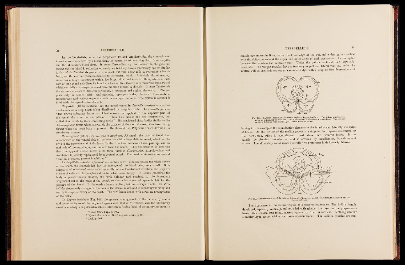
In Lepræa lapidaria (Fig. 148) the general arrangement of the cuticle, hypoderm
and muscular layers of the body-wall agrees with that in P. nebulosa, and the alimentary
canal is similarly slung dorsally, whilst inferiorly a double band of mesentery, apparently
1 ‘Annél. Chét. Nap.,’ p. 399.
8 ‘Quart. Joum. Micr. Sci./ N.s., vol. xxviii, p. 261.
3 Ibid., p. 264.
containing contractile Bbres, leaves the lower edge of the gut* and widening, is attached
with the oblique muscle at the upper and outer angle of each nerve-area. In the space
between the bands is die ventral vessel. Under the gut on each side is a large sub-
intestinal. The oblique muscles have a tendency to pull the lateral wall and make the
ventral wall 'on each side project as a rounded ridge with a deep median depression, and
Fm 148.—Transverse section of the anterior region of cause an indentation on each side. The lower folds Loefp trhteea i nlatpesidtianreia a. re pTrhoem oibnleinqtu.e mTuhsec clecse,l o©mmi.c, cavity is distended with sperms (spm.); other letters as before.
having in ripe examples the reproductive elements in the interior and laterally the ridge
of hooks. At the bottom of the median groove is a ridge in the preparations containing
the nerve-area, which is cone-shaped, broad above and pointed below. It lies
outside the circular muscular coat and is covered by neurilemma, hypoderm and
cuticle. The alimentary canal shows ventrally two prominent folds like a typhlosole.
Pig. 149.—Transverse section of the anterior boLdeyt-twerasl la osf b Peofolyrec.irrus aurantiacus, Grube, in the line of the feet.
The hypoderm in the anterior region of Polycirrus awrawtiacus (Fig. 149) is largely
developed, especially ventrally, and orowded with glands, this layer in the preparations
being often thrown into foliate masses apparently from its softness. A strong circular
muscular layer occurs within the basement-membrane. The oblique muscles are con