28 SABELLARTA ALVEOLATA.
the filaments spring from the summit as a single row. The oèntral axis is deeply stained
by methylene-blue (Arnold Watson).
In spirit the buccal region still retains deep purplish-
brown pigment on the sides, especially external to the
tentacles and between their basal folds. It is terminated
on each side posteriorly, as in S. spinulosa, by a fillet which
has a branchia dorsally and a flattened and pointed lamella
with a minute bristle-tuft below it ventrally, the bristles
having proportionally stout shafts and tapering, closely
spinous tops. They appear to belong to the ventral
series.
The long dorsal bristles in the complex region of
Sabellaria spinulosa are thus absent, only developing paleas
occurring towards its anterior margin. Whether the smooth
bristles to the exterior of the ventral mouth-lobes represent
their equivalent or otherwise is at present unknown, but
their absence dorsally is noteworthy. Meyer’s view that
such represents the dorsal division of the first segment
of the body is thus not without basis, more especially as
the tuft of characteristic bristles shows that the ventral
division is also present. The dorsal bristles seem to be
well developed in Tetreres murata, Allen1; the first branchia
with the fillet and the papilla on its anterior margin would
thus appear to complete the parts of "the first bristled
segment.
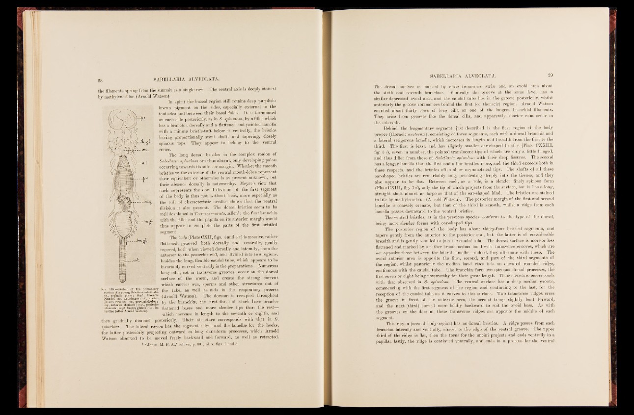
The body (Plate CXII, figs. 4 and 4 a) is massive, rather
flattened, grooved both dorsally and ventrally, gently
tapered, both when viewed dorsally and laterally, from the
anterior to the posterior end, and divided into two regions,
besides the long, flexible caudal tube, which appears to be
invariably curved ventrally in the preparations. Numerous
long cilia, set in transverse grooves, occur on the dorsal
surface of the worm, and create the strong current
which carries ova, sperms and other structures out of
Fig. 139.—Sketch of the alimentary , j well as aids system of a young SabeUana alveolata. » in the respr ir- atordy Aprocess
pc., cephalic plate; th.gi.,^ thoracic (Arnold Watson). The dorsum is occupied throughout
by the branchiae, the first three of which have broader
flattened bases and more slender tips than the rest—
which increase in length to the seventh or eighth, and
gglearnoduss ;l amase.l,l aoee ;s oppvh.,a gpurso ;v eunlt.,r iouuncluinsi ;- mstgo.m, aancthe ;r iborr. gsl.t,o bmroawchn ; gmlagn'd.,s p ; oi-s*te rior testine (after Arnold "Watson).
then gradually diminish posteriorly. Their structure corresponds with that in 8.
spinulosa. The lateral region has the segment-ridges and the lamellae for the hooks,
the latter posteriorly projecting outward as long cuneiform processes, which Arnold
Watson observed to be moved freely backward and forward, as well as retracted.
1 ' Journ. M. B. A.,’ vól. vii, p. 801, pi. x, figs. 1 and 8.
The dorsal surface is marked by close transverse strias and an ovoid area about
the sixth and seventh branchige. Ventrally the groove at the same level has a
similar depressed ovoid area, and the caudal tube lies in the groove posteriorly, whilst
anteriorly the groove commences behind the first (or thoracic) region. Arnold Watson
counted about thirty rows of long cilia on one of the longest branchial filaments.
They arise from grooves like the dorsal cilia, and apparently shorter cilia occur in
the intervals.
Behind the fragmentary segment just described is the first region of the body
proper (thoracic auctonom), consisting of three segments, each with a dorsal branchia and
a lateral setigerous lamella, which increases in length and breadth from the first to the
third. The first is least, and has slightly smaller oar-shaped bristles (Plate CXXIII,
fig. 8 c), seven in number, the pointed translucent tips of which are only a little fringed,
and thus differ from those of Sabellaria spinulosa with their deep fissures. The second
has a longer lamella than the first and a few bristles more, and the third exceeds both in
these respects, and the bristles often show asymmetrical tips. The shafts of all these
oar-shaped bristles are remarkably long; penetrating deeply into the tissues, and they
also appear to be flat. Between each, as a rule, is a slender finely spinous form
(Plate CXIII, fig. 8 d), only the tip of which projects from the surface, but it has a long,
straight shaft almost as large as that of the oar-shaped kind. The bristles are stained
in life by methylene-blue (Arnold Watson). The posterior margin of the first and second
lamellge is coarsely crenate, but that of -the third is smooth, whilst a ridge from each
lamella passes downward to 'the ventral bristles.
The ventral bristles, as in the previous species, conform to the type of the dorsal,
being more slender forms with oar-shaped tips.
The posterior region of the body has about thirty-four bristled segments, and
tapers gently from the anterior to the posterior end, but the latter is of considerable
breadth and is gently rounded to join the caudal tube. The dorsal surface is more or less
flattened and marked by a rather broad median band with transverse grooves, which are
not opposite those between the lateral lamellae—indeed, they alternate with these. The
ovoid anterior area is opposite the first, second, and part of the third segments of
the region, whilst posteriorly the median band rises into an elevated rounded ridge,
continuous with the caudal tube. The branchias form conspicuous dorsal processes, the
first seven or eight being noteworthy for their great length. Their structure corresponds
with that observed in S. spinulosa. The ventral surface has a deep median groove,
commencing with the first segment of the region and continuing to the last, for the
reception of the caudal tube as it curves to this surface. Two transverse ridges cross
the groove in front of the anterior area, the second being slightly bent forward,
and the next (third) curved more boldly backward to suit the ovoid boss. As with
the grooves on the dorsum, these transverse ridges are opposite the middle of each
segment.
This region (second body-region) has no dorsal bristles. A ridge passes from each
branchia laterally and ventrally, almost to the edge of the ventral groove. The upper
third of the ridge is flat, then the torus for the uncini projects and ends ventrally in a
papilla; lastly, the ridge is continued ventrally, and ends in a process for the ventral