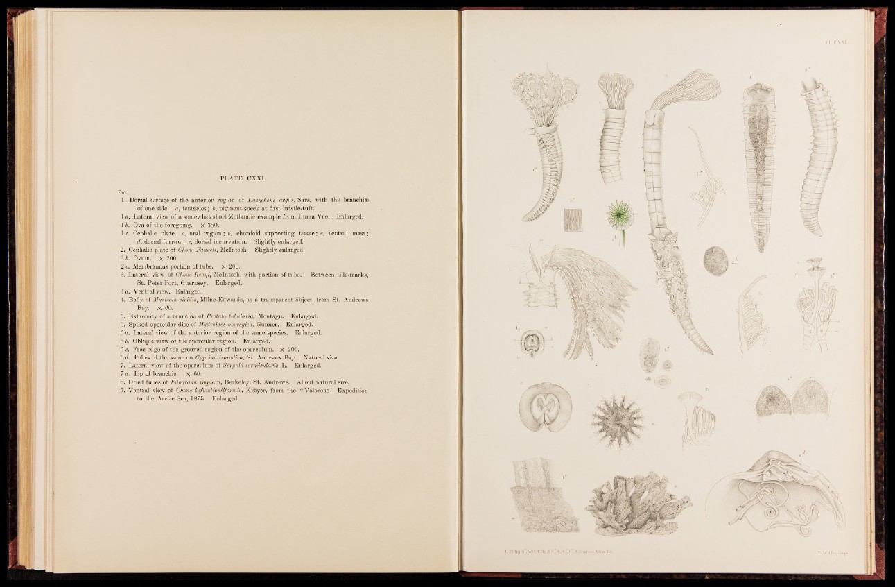
PLATE CXXI.
Fig.
1. Dorsal surface of the anterior region of Dasychone argus, Sars, with the branchiae
of one side, a, tentacles; b, pigment-speck at first bristle-tuft.
1 a. Lateral view of a somewhat short Zetlandic example from Burra Voe. Enlarged.
1 b. Ova of the foregoing, x 350.
1 c. Cephalic plate, a, oral region; b, chordoid supporting tissue; c, central mass;
d, dorsal furrow; e, dorsal incurvation. Slightly enlarged.
2. Cephalic plate of Glione Fauveli, McIntosh. Slightly enlarged.
2 b. Ovum. X 200.
2 c. Membranous portion of tube. X 200.
3. Lateral view of Ghone Reayi, McIntosh, with portion of tube. Between tide-marks,
St. Peter Port, Guernsey. Enlarged.
3 a. Ventral view. Enlarged.
4. Body of Myxioola viridis, Milne-Edwards, as a transparent object, from St. Andrews
Bay. X 60.
5. Extremity of a branchia of Protnla tubularia, Montagu. Enlarged.
6. Spiked opercular disc of Hydroides norvegica, Gunner. Enlarged.
6 a. Lateral view of the anterior region of the same species. Enlarged.
6 6. Oblique view of the opercular region. Enlarged.
6 c. Free edge of the grooved region of the operculum. X 200.
6 d. Tubes of the same on Gyprina islandica, St. Andrews Bay. Natural size.
7. Lateral view of the operculum of Serpula vermicularis, L. Enlarged.
7 a. Tip of branchia. X 60.
8. Dried tubes of Filograna implexa, Berkeley, St. Andrews. About natural size.
9. Ventral view of Ghone infu/ndibuliformis, Kroyer, from the “ Valorous ” Expedition
to the Arctic Sea, 1875. Enlarged.