In the Dodo, thè hind toe is about one third shòrter than the inner, which, as in all
strictly ground Pigeons, is distinctly longer than the outer ; and the middle digit is not
much longer than either of the lateral toes, but it is shorter than the metatarsus. The ungual
joint of the outer toe only is preserved; the others are carefully restored in Plate XII.
Fig. 1, 1 a, 1 by as to length, from the foot covered with integuments, in the British
Museum.1 The phalanges have the usual form, and hence, it is unnecessary to enter into
detail ; the metatarsal articular surface of the first joint of each toe is seen in Plate XII.
Fig. 2. The proximal {ib. Fig. 4,) and distal {ib. Fig. 4 a,) articular facets of one of the
intermediate joints, viz., the second of the outer toe, are also figured.
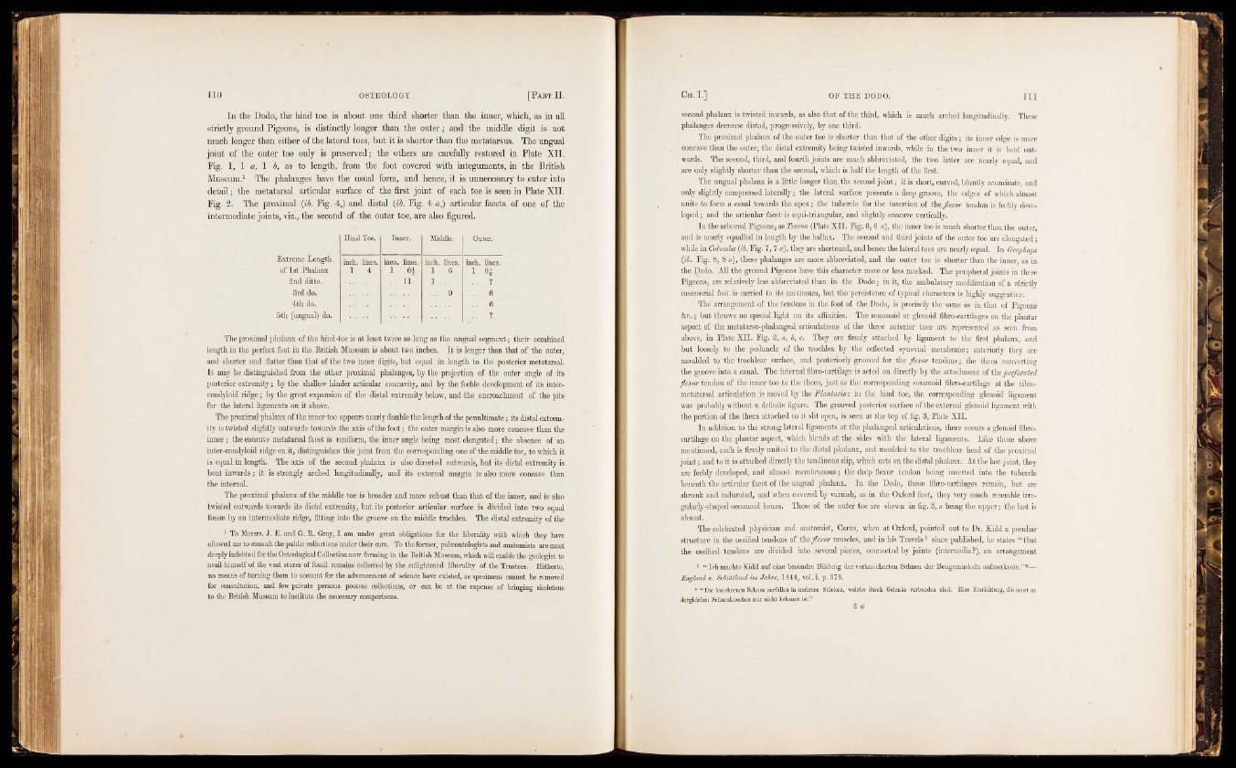
Hind Toe. Inner. Middle. Outer.
Extreme Length inch, lines. inco. lines. inch, lines. inch, lines.
of 1st Phalanx 1 4 1 1 6 1 H
2nd ditto. . . 11 1 . . .. 7
3rd do. 9 .. 6
4th da. .. 6
5th (ungual) do. .. 7
The proximal phalanx of the hind-toe is at least twice as long as the ungual segment; their combined
length in the perfect foot in the British Museum is about two inches. It is longer than that of the outer,
and shorter and flatter than that of the two inner digits, but equal in length to the posterior metatarsal.
It may be distinguished from the other proximal phalanges, by the projection of the outer angle of its
posterior extremity; by the shallow hinder articular concavity, and by the feeble development of its inter-
condyloid ridge; by the great expansion o f the distal extremity below, and the encroachment of the pits
for the lateral ligaments on it above.
The proximal phalanx of the inner toe appears nearly double the length of the penultimate; its distal extremity
is twisted slightly outwards towards the axis of the foot; the outer margin is also more concave than the
inner; the concave metatarsal facet is reniform, the inner angle being most elongated; the absence of an
inter-condyloid ridge on it, distinguishes this joint from the corresponding one of the middle toe, to which it
is equal in length. The axis of the second phalanx is also directed outwards, but its distal extremity is
bent inwards; it is strongly arched longitudinally, and its external margin is also more concave than
the internal.
The proximal phalanx of the middle toe is broader and more robust than that of the inner, and is also
twisted outw.ards towards its distal extremity, but its posterior articular surface is divided into two equal
fossae by an intermediate ridge, fitting into the groove on the middle trochlea. The distal extremity of the
1 To Messrs. J. E. and G. R. Gray, I am under great obligations for the liberality with which they have
allowed me to consult the public collections under their care. To the former, paleontologists and anatomists are most
deeply indebted for the Osteological Collection now forming in the British Museum, which will enable the geologist to
avail himself of the vast stores of fossil remains collected by the enlightened liberality of the Trustees. Hitherto,
no means of turning them to account for the advancement of science have existed, as specimens cannot be removed
for consultation, and few private persons possess collections, or can be at the expense of bringing skeletons
to the British Museum to institute the necessary comparisons.
second phalanx is twisted inwards, äs also that of the third, which is much arched longitudinally. These
phalanges decrease distad, progressively, by one third.
The proximal phalanx of the outer toe is shorter than that of the other digits ; its inner edge is more
concave than the outer, the distal extremity being twisted inwards, while- in the two inner it is bent outwards.
Thé second, third, and fourth joints are much abbreviated, the two latter are nearly equal, and
are only slightly shorter than the second, which is half the length of the first.
The ungual phalanx is a little longer than the second joint; it is short, curved, bluntly acuminate, and
only slightly compressed laterally ; the lateral surface presents a deep groove, the edges of which almost
unite to form a canal towards the apex; the tubercle for the insertion of th§ flexor tendon is feebly developed
; and the articular facet is equi-triangular, and slightly concave vertically.
In the arboreal Pigeons, as Treron (Plate XII. Fig. 6 ,6 a), the inner toe is much shorter than the outer,
and is nearly equalled in length by the hallux. The second and third joints of the outer toe are elongated •
while in Columba {ib. Fig. 7 ,7 a), they are shortened, and hence the lateral toes are nearly equal. In Geophaps
{ib. Fig. 8, 8 a), these phalanges are more abbreviated, and the outer toe is shorter t.hau the inner, as in
the Dodo. All the ground Pigeons have this character more or less marked. The peripheral joints in these
Pigeons, are relatively less abbreviated than in the Dodo ; in it, the ambulatory modification of a strictly
insessorial foot is carried to its maximum, but the persistence of typical characters is highly suggestive.
The arrangement of the tendons in the foot of the Dodo, is precisely the same as in that of Pigeons
&e. ; but throws no special light on its affinities. The sesamoid or glenoid fibro-cartilages on the plantar
aspect of the metatarso-phalangeal articulations of the three anterior toes are represented as seen from
above, in Plate XII. Fig. 2, a, b, c. They are firmly attached by ligament to the first phalanx, and
but loosely to the peduncle of the trochlea by the reflected synovial membrane; anteriorly they are
moulded to the trochlear surface, and posteriorly grooved for the flexor tendons ; the theca converting
the groove into a canal. The internal fibro-cartilage is acted on directly by the attachment of the perforated
flexor tendon of the inner toe to the theca, just as the corresponding sesamoid fibro-cartilage at the tibio-
nietatarsal articulation is moved by the Plantaris : in the hind toe, the corresponding glenoid ligament
was probably without a definite figure. The grooved posterior surface of the external glenoid ligament with
the portion of the theca attached to it slit open, is seen at the top of fig. 3, Plate XII.
In addition to the strong lateral ligaments at the phalangeal articulations, there occurs a glenoid fibro-
cartilage on the plantar aspect, which blends at the sides with the lateral ligaments. Like those above
mentioned, each is firmly united to the distal phalanx, and moulded to the trochlear head of the proximal
joint ; and to it is attached directly the tendinous slip, which acts on the distal phalanx. At the last joint, they
are feebly developed, and almost membranous ; the deep flexor tendon being inserted into the tubercle
beneath the articular facet of the ungual phalanx. In the Dodo, these fibro-cartilages remain, but are
shrunk and indurated, and when covered by varnish, as in the Oxford foot, they very much resemble irregularly
shaped sesamoid bones. Those of the outer toe are shewn in fig..3, a being the upper; the last is
absent.
The celebrated physician and anatomist, Carus, when at Oxford, pointed out to Dr. Kidd a peculiar
structure in the ossified tendons of the flexor muscles, and in his Travels1 since published, he states “ that
the ossified tendons are divided into several pieces, connected by joints (intemodia ?}, an arrangement
1 “ Ich machte Kidd auf eine besondre Bildung der verknöcherten Sehnen der Beugemuskeln aufmerksam.”*—
England u. Schottland im Jahre, 1844, vol. i. p. 375.
* “ Die knöchernen Sehnen zerfallen in mehrere Stücken, welche durch Gelenke verbunden sind. Eine Einrichtung, die sonst an
dergleichen Sehnenknochen mir nicht bekannt ist.”
2 G