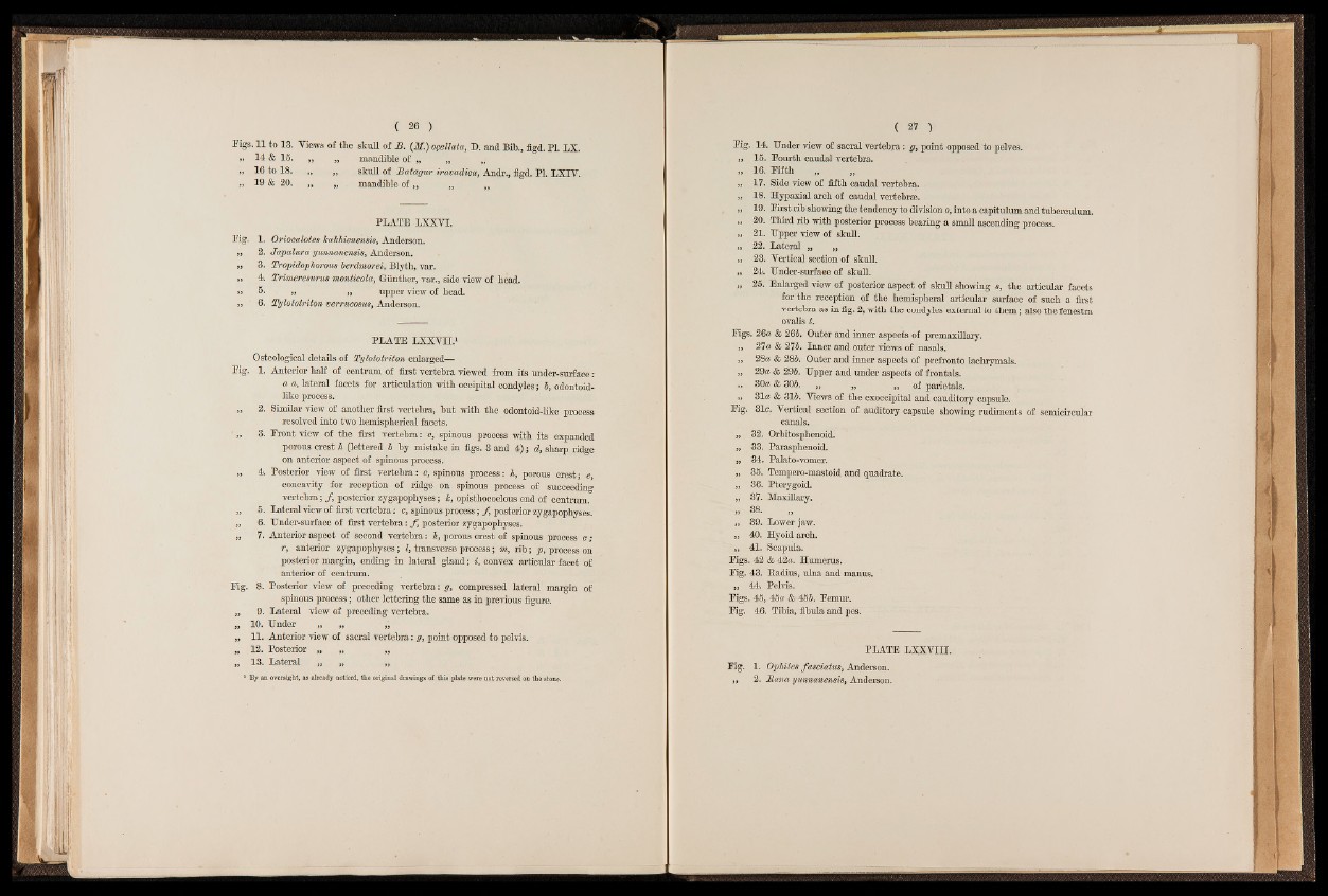
Figs. 11 to 13. Views of the skull of B. (iff.) ogellata, D. and Bib., figd. PI. LX.
„ 14 & 15. „ „ mandible of „ „ „
„ 16 to 18. „ „ skull of Batagur iravadica, Andr., figd. PI. LXIV.
„ 19 & 20. ,, „ . mandible of „ „ „
PLATE LXXVI.
Eig. 1. Oriocalotes kakhienensis, Anderson.
,, 2. Japalura yimnaneims, Anderson.
„ 3. Tropidophorous berdmorei, Blyth, yar.
„ 4. Trvmeresurus monticola, Günther, var., side view of bead.
>» 5* » „ upper view of bead.
„ 6. Tylototriton verrucosus, Anderson.
PLATE LXXVII.1
Osteológica! details of Tylototriton enlarged—
Eig. 1. Anterior half of centrum of first vertebra viewed from its under-surface:
a a, lateral facets for articulation with occipital condyles; 5, odontoid-
like process.
„ 2. Similar view of another first vertebra, but with the odontoid-like process
resolved into two hemispherical facets.
' „ 3. Eront view of the first vertebra : c, spinous process with its expanded
porous crest h (lettered b by mistake in figs. 3 and 4); d, sharp ridge
on anterior aspect of spinous process.
„ 4. Posterior view of first vertebra: c, spinous process: h, porous crest; e,
concavity for reception of ridge on spinous process of succeeding
vertebra; / , posterior zygapopbyses; k, opistbocoelous end of centrum.
„ 5. Lateral view of first vertebra: c, spinous process; f posterior zygapopbyses.
„ 6. Under-surface of first vertebra: f posterior zygapopbyses.
,, 7. Anterior aspect of second vertebra: h, porous crest of spinous process c ;
r, anterior zygapopbyses; I, transverse process; m, rib; p, process on
posterior margin, ending in lateral gland; i, convex articular facet of
anterior of centrum.
Eig. 8. Posterior view of preceding vertebra: g, compressed lateral margin of
spinous process; other lettering the same as in previous figure.
,, 9. Lateral view of preceding vertebra.
„ 10. Under „ „
„ 11. Anterior view of sacral vertebra: g, point opposed to pelvis.
„ 12. Posterior „ „ „
„ 13. Lateral „ „ ,,
1 By an oversight, as already noticed, the original drawings of this plate were not reversed on the stone.
Kg. 14 Under view of sacral vertebra: g, point opposed to pelves.
„ 15. Eourth caudal vertebra.
„ 16. Eiftb
„ 17. Side view of fifth caudal vertebra.
„ 18. Hypaxial arch of caudal vertebrae.
„ 19. Eirst rib showing the tendency to division o, into a capitulum and tuberculum.
,, 20. Third rib with posterior process bearing a small ascending process.
„ 21. Upper view of skull.
„ 22. Lateral „ „
„ 23. Vertical section of skull.
„ 24. Under-surfaee of skull.
„ 25. Enlarged view of posterior aspect of skull showing s, the articular facets
for the reception of the hemispheral articular surface of such a firstvertebra
as in fig. 2, with the condyles external to them; also the fenestra
ovalis t.
Eigs. 26a & 26b. Outer and inner aspects of premaxillary.
,, 27a & 2tJb. Inner and outer views of nasals.
,, 28« & 285. Outer and inner aspects of prefronto lachrymals.
,, 29« & 295. Upper and under aspects of frontals.
„ 30« & 305. „ ,j, „ of parietals.
,, 31« & 315. Views of the exoccipital and cauditory capsule.
Eig. 31c. Vertical section of auditory capsule showing rudiments of semicircular
canals.
,, 32. Orbitosphenoid.
,, 33. Parasphenoid.
,, 34. Palato-vomer.
„ 35. Tempero-mastoid and quadrate.
„ 36. Pterygoid.
,, 37. Maxillary.
„ 38.
„ 39. Lower jaw.
„ 40. Hyoid arch.
41. Scapula.
Eigs. 42 & 42«. Humerus.
Eig. 43. Radius, ulna and manus.
„ 44. Pelvis.
Eigs. 45, 45« & 455. Eemur.
Eig. 46. Tibia, fibula and pes.
PLATE LXXVIII.
Fig. 1. Ophites fasciatus, Anderson.
,, 2. Bana yimnanensis, Anderson.