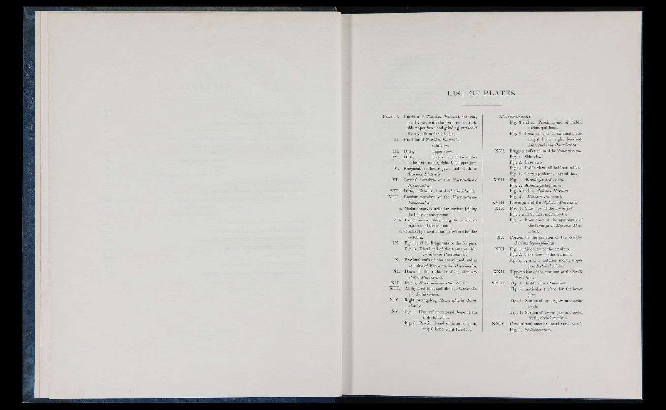
LIST OF PLATES.
II.
II I.
IV .
VI.
VII.
V III.
Cranium of Toxodon Platensis, iiat. size,
basal view, with the sixth molar, right
side upper jaw, and grinding surface of
the seventh molar left side.
Cranium of Toxodon Platensis,
side view.
Ditto, upper view.
Ditto, back view, withtwo views
of the sixth molar, right side, upper jaw.
Fragment of lower jaw, and teeth of
Toxodon Platensis.
Cervical vertebrfe of the Macraiichenia
Patachonica.
Ditto, ditto, and of Auchenia Llama.
Lumbar vertehree of the Macrauchenia
Patachonica.
a. Medium convex articular surface joining
the body of the sacrum.
h. b. Lateral concavities joining the transverse
processes of the sacrum,
c. Ossified ligament of an auchyloscd lumbar
vertebra.
Fig. 1 and 2. Fragments of the Scapula.
Fig. 3. Distal end of tlie femur of Macrauchenia
Patachonica.
Proximal ends of the anchylosod radius
and ulna of Macrauchenia Patachonica.
X I. Bones of the right fore-foot, Macrauchenia
Patachonica.
Femur, Macrauchenia Patachonica.
Anchylosod tibia and fibula, Macrauchenia
Patachonica.
Right astragalus, Macrauchenia Patachonica,
X V . Fig. 1. External metatarsal bone of the
right hind-foot.
Fig. 2. Proximal end of internal metacarpal
bone, right fore-foot.
IX.
X.
X I I.
X I II.
XIV.
XV. {continued.')
Fig. 3 and 4. Proximal end of middle
metacarpal bone.
Fig. 5 Proximal end of externa! metacarpal
bone, right fore-foot,
Macrauchenia Patachonica.
X VI. Fragment of cranium oftheCYoisoMeri«?«.
Fig. 1. Side view.
Fig. 2. Base view.
Fig. 3. Inside view, all half natural size.
Fig. 4. Os tyinpanicum, natural size.
X V II. F ig .] . Megalonyx Jcffcrsonii.
Fig. 2. Megalonyx laqueatus.
Fig. 3 and 4-. Mylodon JIarlani.
Fig. 5. Mylodon Daricinii.
X V III. Lower jaw of the Mylodon Darwinii.
X IX . Fig. 1. Side view of the lower jaw.
Fig. 2 and 3. La.st molar tooth.
Fig. 4. Front view of the symphysis of
the lower jaw, Mylodon Dar-
vcxnii.
X X . Portion of the skeleton of the Scelido-
therium leptocephalum.
X X I. Fig. 1. Side view of the cranium.
Fig. 2. Back view of the cranium.
Fig. 3, 4, and 5, anterior molar, upper
jaw Scelidotherium.
X X I I, Upper view of the cranium of the Scelidotherium.
X X I II. Fig. 1. Inside view of cranium.
Fig. 2. Articular surface for the lower
jaw.
Fig. 3. Section of upper jaw and molar
teeth.
Fig. 4. Section of lower jaw and molar
teeth, Scelidotherium.
X X IV . Cervical and anterior dorsal vertebr« of,
Fig. 1. Scelidotherium.