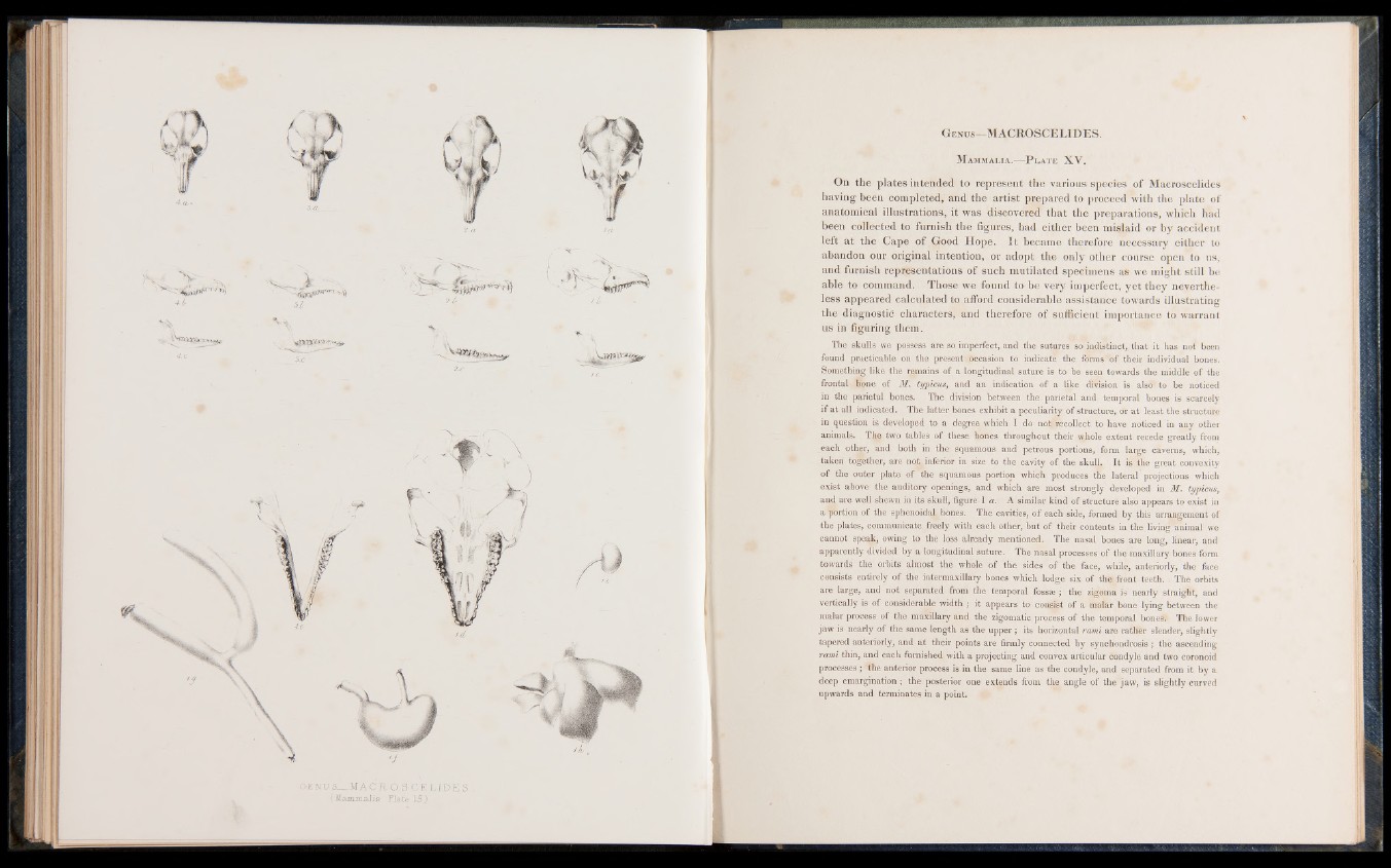
GENUS__MA C R O S CE LII7E S .
( Mammalia Plate. 15)
G en u s— MACROSCELIDES.
M ammalia.— P l a t e XV.
On the plates intended to represent the various species of Macroscelides
having been completed, and the artist prepared to proceed with the plate of
anatomical illustrations, it was discovered that the preparations, which had
been collected to furnish the figures, had either been mislaid or by accident
left at the Cape of Good Hope. It became therefore necessary either to
abandon our original intention, or adopt the only other course open to us,
and furnish representations of such mutilated specimens as we might still be
able to command. Those we found to be very imperfect, yet they nevertheless
appeared calculated to afford considerable assistance towards illustrating
the diagnostic characters, and therefore of sufficient importance to warrant
us in figuring them.
The skulls we possess are so imperfect, and the sutures so indistinct, that it has not been
found practicable on the present Occasion to indicate the forms of their individual bones.
Something like the remains of a longitudinal suture is to be seen towards the middle of the
frontal lone of M. typicus, and an indication of a like division is also to be noticed
in the parietal bones. The division between the parietal and temporal bones is scarcely
if at a.11 indicated. The latter bones exhibit a peculiarity of structure, or at least the structure
in question is developed to a degree which I do no|jfrecollect to have noticed in any other
animals. The two tables of these bones throughout their whole extent recede greatly from
each otlmr, and both in the squamous and petrous portions, form large caverns, which,
taken together, are not inferior in size to the cavity of the skull. It is the great convexity
of the outer plate of the squamous portion which produces the lateral projections which
exist above the auditory openings, and which are most strongly developed in M. typicus,
and are well shewn in its skull, figure l a . A similar kind of structure also appears to exist in
a portion of the sphenoidal; bones. The cavities, of each side, formed by this arrangement of
the plates, communicate freely with each other, but of their contents in the living animal we
cannot speak, owing to the loss already mentioned. The nasal bones are long, linear, and
apparently divided by a longitudinal suture. The nasal processes of the maxillary bones form
towards the; orbits almost the whole of the sides of the face, while, anteriorly, the face
consists entirely of the intermaxillary bones which lodge six of the front teeth. . The orbits
are large, and not separated from the temporal fossae ; the zigoma is nearly straight, and
vertically is of considerable width ; it appears to consist of a malar bone lying between the
malar process of the maxillary and the zigomatic process of the temporal bones; The lower
jaw is nearly of the same length as the upper; its horizontal rami are rathfer slender, slightly
tapered anteriorly, and at their points are firmly connected by synchondrosis ; the ascending
rami thin, and each furnished with a projecting and convex articular condyle and two coronoid
processes; the anterior process is in the same line as the condyle, and separated from it by a
deep emargination; the posterior one extends from the angle of the jaw, is slightly curved
upwards and terminates in a point.